Acromioclavicular Joint Injury
Treatment of acromioclavicular ligament injury and AC joint arthritis
Overview
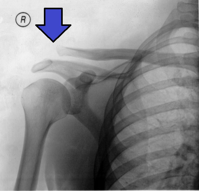
An acromioclavicular (AC) joint injury is a sprain or rupture of the ligaments of the joint between the outer end of the collarbone and the acromion of the shoulder blade. Much of what is popularly called a dislocated shoulder is in fact an injury of this joint, which is quite different from a true shoulder dislocation. It usually follows a direct fall onto the point of the shoulder and is common in cycling, motorcycling, skiing, football and rugby. The great majority are treated without surgery; operation is considered for severe separations and in selected athletes. The same joint can also become painful through wear over the years (AC joint arthritis).
Anatomy of the AC Joint and Mechanism of Injury
The collarbone is the only bony connection between the trunk and the arm. Its outer end forms a small joint with the acromion of the shoulder blade, and two groups of ligaments provide the stability of this joint.
The acromioclavicular ligaments surround the joint itself and control front-to-back stability. The coracoclavicular ligaments (conoid and trapezoid) connect the collarbone to the coracoid process and act as the principal suspension in the vertical direction.
Injury typically occurs with a fall onto the point of the shoulder with the arm at the side. The blow drives the acromion downwards while the collarbone stays in place, and the ligaments fail in sequence: first the acromioclavicular ligaments, then, if force continues, the coracoclavicular ligaments.
When the coracoclavicular ligaments rupture, the outer end of the collarbone stands proud under the skin. In reality it is not the collarbone rising but the shoulder dropping — a distinction that explains the logic of treatment.
AC joint arthritis is a separate condition, caused by repetitive loading and age-related wear. It is common in weightlifters and those who bench press or train overhead, and causes pain and tenderness at the outer end of the collarbone.
The Rockwood Classification
- Type 1: sprain of the acromioclavicular ligaments without rupture. No displacement on radiographs
- Type 2: the acromioclavicular ligaments are ruptured, the coracoclavicular ligaments intact, with partial displacement
- Type 3: both ligament groups are ruptured and the collarbone is clearly elevated. This is the type whose treatment is most debated. We prefer surgery in athletes and active young patients; the indication is always set by the patient rather than by the injury type, weighing age, occupation, level of sport, expectations and the functional demand on the shoulder
- Type 4: the outer end of the collarbone is displaced backwards into the trapezius muscle. Requires surgery
- Type 5: a severe form of type 3, with marked prominence and stripping of the muscle attachments. Requires surgery
- Type 6: a very rare type in which the collarbone is displaced beneath the acromion or coracoid. Requires surgery
Symptoms
- Sharp pain and marked tenderness at the point of the shoulder, over the outer end of the collarbone
- A visible prominence and change in shoulder contour, more obvious with greater separation
- Pain on bringing the arm across the body towards the opposite shoulder
- Difficulty raising the arm overhead and lying on that side
- Piano key sign: the elevated end of the collarbone can be pushed down and springs back up on release
- A sense of the arm being dragged down and the shoulder tiring when carrying weight
- In AC joint arthritis: insidious pain aggravated by bench press and overhead work, with point tenderness over the joint
Making the Diagnosis
The diagnosis is usually clinical. Point tenderness over the joint, a visible prominence and the piano key sign are decisive. The cross-body adduction test reproduces pain arising from the AC joint.
Plain radiographs are the standard investigation. Alongside a standard shoulder view, dedicated views demonstrate the AC joint, and comparison views of both shoulders allow the degree of separation to be measured. An axillary view is necessary to identify a type 4 injury.
Weighted stress views are no longer used routinely; they increase pain and rarely alter the treatment decision.
MRI is obtained to identify associated rotator cuff and labral injury, particularly after high-energy trauma and in athletes. In AC joint arthritis, MRI shows joint oedema, but this finding is also common in people without symptoms and must be interpreted with the examination.
Non-operative Treatment
Almost all type 1 and type 2 injuries, and the majority of type 3 injuries, are treated successfully without surgery. The contour change may be permanent, but shoulder strength and function return fully in most patients.
- Sling: usually 1 - 3 weeks until pain settles, giving early comfort
- Ice and anti-inflammatory medication
- Early movement: pendulum and passive exercises within pain limits to prevent stiffness
- Graded strengthening of the deltoid, trapezius and scapular muscles to distribute load
- Temporary avoidance of heavy lifting and contact sport, usually for 6 - 12 weeks
- In AC joint arthritis: activity modification (limiting the range of bench press and dips), anti-inflammatory treatment and intra-articular corticosteroid injection
- In type 3 injuries non-operative treatment is tried first; if symptoms persist, later surgery gives results comparable to early surgery
Surgery & First 3 Weeks
The aim of surgery is to re-suspend the collarbone from the coracoid process, restoring the anatomical relationship and allowing the ligaments to heal. Techniques differ between acute and chronic injuries: in chronic cases the ligaments have lost their capacity to heal, and reconstruction with a tendon graft is required.
Indications for Surgery
- Type 4, 5 and 6 injuries: surgery is indicated
- Type 3 injuries: surgery is preferred in athletes, active young patients, manual workers and overhead workers; the decision follows the patient, not the injury type
- Pain and weakness persisting 3 - 6 months despite non-operative treatment
- An associated collarbone fracture or rotator cuff tear
- In isolated AC joint arthritis: pain unresponsive to injection and activity modification
- Surgery is not undertaken for appearance alone, as it carries its own risks and leaves a scar
Coracoclavicular Stabilisation in Acute Injury
Within the first 3 - 6 weeks the ruptured ligaments can still be expected to heal. High-strength suture and button (endobutton) constructs are placed between the collarbone and the coracoid process to hold the bone in its anatomical position.
The procedure can be performed arthroscopically assisted or open. Arthroscopic assistance allows safe visualisation of the undersurface of the coracoid and assessment of associated intra-articular pathology at the same sitting.
Healing here occurs in the patient's own ligaments; the implant provides temporary support while that happens.
Ligament Reconstruction in Chronic Injury
Once months have passed, the ligaments have lost their healing capacity. In these cases the coracoclavicular ligaments are reconstructed with a tendon graft, usually a hamstring autograft or an allograft.
The graft is passed around the coracoid process and fixed to the collarbone, creating a biological suspension. Suture and button constructs alone are insufficient in chronic cases and carry a high risk of loss of reduction.
Distal Clavicle Excision
In AC joint arthritis and where pain arises within the joint itself, a few millimetres of the outer end of the collarbone are removed to eliminate the painful contact.
This can be done arthroscopically and rehabilitates quickly. The amount removed matters: too little leaves persistent pain, too much destabilises the joint. The coracoclavicular ligaments must be preserved.
The Operation
- General anaesthesia, often with a nerve block
- Operating time varies from 45 to 120 minutes depending on technique
- One overnight stay suffices in most cases; distal clavicle excision can be a day case
- A sling is usually worn for 4 - 6 weeks after stabilisation, and this period is not shortened
- Follow-up radiographs confirm that the reduction has been maintained
Risks & Complications
The main problem after AC joint surgery is loss of reduction — the collarbone rising again during healing. No technique eliminates this risk entirely, and the patient's adherence to postoperative restrictions directly affects the outcome.
General Surgical Risks
- Infection
- Bleeding and haematoma
- Risks related to anaesthesia and the nerve block
- Wound healing problems and a visible scar, as the incision lies over the shoulder
- Shoulder stiffness
Risks Specific to This Operation
- Loss of reduction: the commonest problem, appearing as partial re-elevation of the collarbone. Function is usually preserved, but the contour changes
- Fracture of the coracoid or clavicle: can occur through the drill tunnels, particularly with early loading
- Implant-related problems: button migration, suture failure, widening of the bone tunnels
- Persistent AC joint pain: degeneration within the joint can cause ongoing pain despite stable fixation, sometimes requiring distal clavicle excision
- Instability from excessive distal clavicle excision
- Graft-related problems: donor site pain, graft stretching
- Heterotopic ossification in the coracoclavicular region: usually symptomless
- Need for further surgery, in cases of lost reduction or implant problems
Rehabilitation
Rehabilitation aims to protect the repaired or reconstructed suspension while preventing shoulder stiffness. Lifting the arm against gravity and carrying weight are restricted early on, because they place a direct distracting force on the fixation. Programmes after non-operative treatment progress more quickly.
Phase 1 — Protection (0 - 6 Weeks)
- Sling, usually for 4 - 6 weeks after surgery, including at night
- Elbow, wrist and finger movement is free
- Passive and assisted movement, with elevation limited to 90 degrees for the first 4 - 6 weeks
- Active elevation of the arm, carrying weight and leaning on the arm are forbidden
- Ice and pain control; stopping smoking directly affects healing
Phase 2 — Restoring Movement (6 - 12 Weeks)
- The sling is discontinued and active movement introduced gradually
- Overhead movement is permitted progressively, aiming for pain-free full range by weeks 10 - 12
- Scapular stabilisation and light isometric strengthening begin
- Resistance work progresses with light bands within pain limits
- Radiographs confirm that the reduction has been maintained
Phase 3 — Strengthening (3 - 5 Months)
- Graded strengthening of the deltoid, trapezius, rotator cuff and scapular muscles
- Graded return to weight training, with bench press and dips — which load the AC joint — left until last and begun with a limited range
- Proprioception and core strengthening
- The goal by the end of this phase is full return to daily and work activities
Return to Work and Sport
- Desk work: 1 - 2 weeks
- Light physical work: 6 - 8 weeks
- Heavy manual and overhead work: 4 - 6 months
- Non-contact sport: usually 3 - 4 months
- Contact and collision sport (football, rugby, judo, competitive cycling and motorcycling): 5 - 6 months
- Criteria for return: pain-free full range of motion, strength at least 90 per cent of the other side, maintained reduction on radiographs and symptom-free performance of sport-specific movements
- After non-operative treatment of type 1 - 2 injuries, return to sport is usually possible within 2 - 6 weeks
Is This Treatment Right for You?
Every patient's condition is different. After a detailed examination and imaging, the treatment plan that suits you best will be prepared.
Would you like more information about Acromioclavicular Joint Injury?
Book Appointment