SLAP Tear
Arthroscopic repair or biceps tenodesis for a tear of the superior labrum at the biceps attachment
Overview
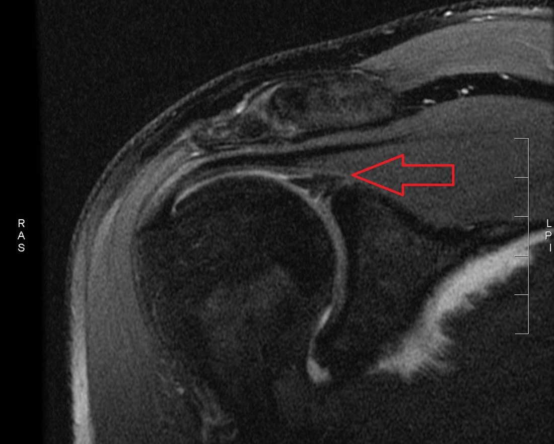
A SLAP tear is a tear of the superior labrum — the cartilage rim of the shoulder socket — running from front to back at the point where the long head of the biceps attaches. It may arise from repeated strain in overhead sports or from a single injury such as a fall on an outstretched arm. Patients describe deep-seated shoulder pain, catching when raising the arm overhead and loss of power. Diagnosis is among the hardest problems in shoulder surgery, because age-related changes at the superior labrum are commonly seen on the scans of people with no symptoms, and a finding on imaging does not by itself call for surgery.
The Labrum, the Biceps Anchor and the Mechanism of Injury
The shoulder is the joint between the ball of the upper arm bone and the shallow socket (glenoid) of the shoulder blade. Because the socket is small relative to the ball, the labrum, a cartilage rim around its edge, deepens the socket and contributes to joint stability while allowing wide movement.
The tendon of the long head of biceps attaches to the superior labrum and the upper rim of the socket; this junction is called the biceps anchor. A SLAP tear involves this region, and the biceps tendon itself must be assessed alongside it.
During the overhead throwing motion, as the arm reaches maximum external rotation, the biceps tendon twists and peels the posterosuperior labrum away. Repeated loading of this kind is the principal mechanism in throwing athletes.
In acute injury the mechanism differs: a fall onto an outstretched hand with the elbow extended, sudden heavy lifting or a sudden traction force on the arm can detach the biceps anchor.
After the age of forty, degenerative change at the superior labrum is common. It is often symptomless, and finding it on imaging is not in itself an indication for treatment.
Types of SLAP Tear
- Type 1: fraying of the superior labral edge with an intact biceps anchor. Usually age-related and does not require repair
- Type 2: detachment of the biceps anchor from the socket — the type that matters most surgically. Subtypes extend anteriorly, posteriorly or in both directions
- Type 3: a bucket-handle tear of the superior labrum that displaces into the joint while the biceps attachment remains intact
- Type 4: a bucket-handle tear extending into the biceps tendon itself; treatment depends on how much of the tendon is involved
- Associated pathology is common: rotator cuff tears, shoulder instability, posterior capsular tightness and biceps tendinitis must all be assessed
Symptoms
- Deep, poorly localised aching in the shoulder: patients typically point inside the joint
- Pain when raising the arm overhead or taking it behind the body
- Catching, locking, clicking or popping, especially with rotation
- In throwing athletes, loss of velocity and control: the so-called dead arm feeling is typical
- Weakness when lifting or pushing
- Night pain: less prominent than in rotator cuff disease
- Tenderness over the biceps groove at the front of the shoulder, suggesting associated biceps tendinitis
Making the Diagnosis
Diagnosis depends on combining history, examination and imaging. The mechanism of injury and, in athletes, the throwing discipline and the effect on performance are explored in detail.
No single examination test is conclusive; tests are interpreted together. The O'Brien active compression test, the biceps load test and the dynamic labral shear test are the most widely used. The rotator cuff, instability and posterior capsular tightness are assessed at the same time; loss of internal rotation frequently accompanies the condition in throwers.
Plain radiographs do not show the labrum and are obtained to exclude arthritis, bony lesions and calcification. Standard MRI lacks sensitivity for superior labral tears. MR arthrography, performed after contrast is injected into the joint, is the most valuable study for demonstrating detachment of the biceps anchor.
A finding on imaging is not sufficient on its own. The diagnosis rests on agreement between the clinical picture and the images; in doubtful cases it is confirmed at arthroscopy.
Non-operative Treatment
Apart from acute, large tears, treatment begins non-operatively. Results are particularly good over the age of forty and where degenerative change predominates.
- Activity modification: a temporary break from overhead and throwing activity
- Anti-inflammatory medication and ice
- Posterior capsular stretching: correcting loss of internal rotation is the cornerstone of treatment in throwers
- Scapular stabilisation training: restoring shoulder blade mechanics reduces load within the joint
- Balanced strengthening of the rotator cuff and scapular muscles
- Review of throwing mechanics: correcting technical faults prevents recurrence in athletes
- Intra-articular corticosteroid injection in selected cases, for pain control and diagnostic information
- Response is assessed after three months of a well-conducted programme
Surgery & First 3 Weeks
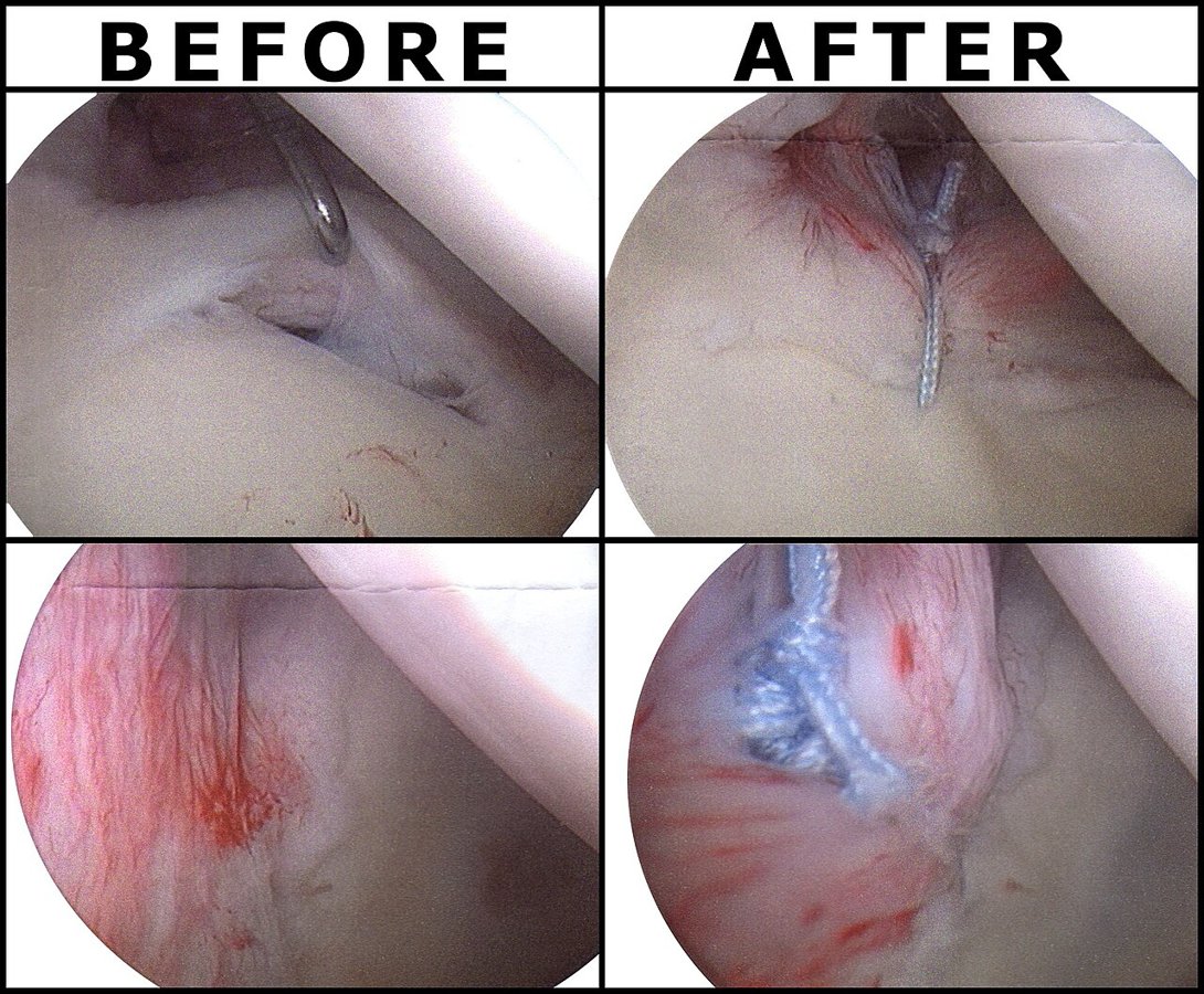
Surgical treatment is performed arthroscopically. The decisive factors are the patient's age, level of sport, the type of tear and the state of the biceps tendon. Repair is favoured in the young throwing athlete, while biceps tenodesis gives more reliable results from middle age onwards.
Indications for Surgery
- Persistent symptoms despite 3 - 6 months of well-conducted non-operative treatment
- Type 2 tears following acute trauma with clear detachment of the biceps anchor
- Bucket-handle tears causing mechanical catching or locking
- Impairment severe enough to prevent an athlete continuing to compete
- Arthroscopy already planned for an associated rotator cuff tear or instability
- An isolated degenerative superior labral finding over the age of forty is not by itself an indication
Arthroscopic Debridement
In type 1 tears and degenerative cases with an intact biceps anchor, the torn and frayed labral tissue is trimmed. The procedure is short, rehabilitation is rapid and joint stability is preserved.
It aims to relieve pain rather than to restore structure, and is insufficient on its own where the biceps anchor is detached.
SLAP Repair
In type 2 tears the detached labrum and biceps anchor are fixed to the upper rim of the socket with suture anchors. The rim is first prepared, then one or two anchors restore the labrum to its anatomical position.
This is preferred in young, active patients and particularly in throwing athletes. Even in this group, however, the rate of full return to previous performance is lower than for most other shoulder operations, and athletes must be counselled realistically before surgery.
Beyond the age of thirty-five to forty, repair carries an increased risk of postoperative stiffness and persistent pain, which is why biceps tenodesis is favoured in this age group.
Biceps Tenodesis and Tenotomy
In biceps tenodesis the long head tendon, the source of the problem, is released from its labral attachment and fixed to the upper arm bone with a screw or suture anchor. This removes the peeling force on the labrum while preserving muscle strength and elbow function.
It is preferred over the age of forty, in patients with associated biceps tendon pathology and after failed SLAP repair. Pain relief and satisfaction are more predictable than with repair in this group.
In tenotomy the tendon is simply released and not fixed. This suits older, lower-demand patients; a bunching of the muscle in the front of the arm (Popeye deformity) and mild cramping may occur.
The Operation
- General anaesthesia, usually with a regional nerve block that markedly reduces pain for the first 12 - 18 hours
- Performed arthroscopically through millimetre portals in the beach-chair or lateral position
- Operating time is about 45 - 90 minutes, longer where associated pathology is treated
- Discharge is usually the same day or after one night
- A sling is worn afterwards, for a period that depends on the procedure performed
Risks & Complications
Arthroscopic shoulder surgery is safe, but SLAP repair has particular drawbacks that must be discussed beforehand — above all that return to previous performance is not guaranteed in the throwing athlete.
General Surgical Risks
- Infection: under 1 per cent in arthroscopic shoulder surgery
- Risks related to anaesthesia and the nerve block
- Injury to vessels or nerves: rare
- Deep vein thrombosis: rare in upper limb surgery
Risks Specific to This Operation
- Postoperative stiffness: the commonest problem after SLAP repair, with increased risk above the age of 35 - 40
- Persistent pain: symptoms may not resolve completely even when the repair heals
- Incomplete return to sport: full return to pre-injury performance is around 60 - 70 per cent in throwing athletes and lower still in very high-demand groups such as professional baseball pitchers
- Suture anchor problems: pull-out, cartilage damage or displacement into the joint
- After biceps tenodesis: pain at the fixation site, rarely failure of fixation and Popeye deformity
- Axillary nerve injury: a rare complication related to portal placement
- Need for further surgery: biceps tenodesis is the usual solution after a failed SLAP repair
Rehabilitation
Rehabilitation differs markedly according to the procedure performed. After SLAP repair, active use of the biceps is restricted early on to protect the healing tissue, whereas programmes after debridement and tenodesis progress faster. The plan below is for SLAP repair.
Phase 1 — Protection (0 - 4 Weeks)
- Sling immobilisation, usually for 3 - 4 weeks, including at night
- Elbow, wrist and finger movement is free
- Supervised passive range of motion is begun, with external rotation kept within neutral limits
- Active biceps contraction and resisted elbow flexion are forbidden: they place a direct peeling force on the repair
- Taking the arm behind the body and forceful stretching are avoided
- Ice and pain control
Phase 2 — Restoring Movement (4 - 8 Weeks)
- The sling is discontinued
- Graded progression from active-assisted to full active movement
- External rotation is increased gradually; posterior capsular stretching begins in throwers
- Scapular stabilisation work and isometric rotator cuff activation
- Light active biceps use without resistance is permitted after week 6
- Goal: pain-free, near-full range of motion by the end of week 8
Phase 3 — Strengthening (8 - 16 Weeks)
- Resistance band strengthening of the rotator cuff and scapular muscles
- Biceps strengthening introduced gradually after weeks 10 - 12
- Closed kinetic chain exercise and core and hip strengthening
- Proprioception and neuromuscular control work
- Overhead lifting is not yet permitted
Phase 4 — Return to Sport (4 - 9 Months)
- Interval throwing programme: distance and intensity increase over weeks, stepping back one stage if pain appears
- Return to contact sport usually at 4 - 6 months
- Full return for overhead and throwing athletes usually at 6 - 9 months
- Criteria for return: pain-free full range of motion, rotator cuff strength at least 90 per cent of the other side, full scapular control and completion of the interval throwing programme without symptoms
- Reviewing throwing mechanics and training load reduces the risk of recurrence
Is This Treatment Right for You?
Every patient's condition is different. After a detailed examination and imaging, the treatment plan that suits you best will be prepared.
Would you like more information about SLAP Tear?
Book Appointment