Proximal Humerus Fracture
Surgical treatment of proximal humerus, clavicle and scapula fractures
Overview
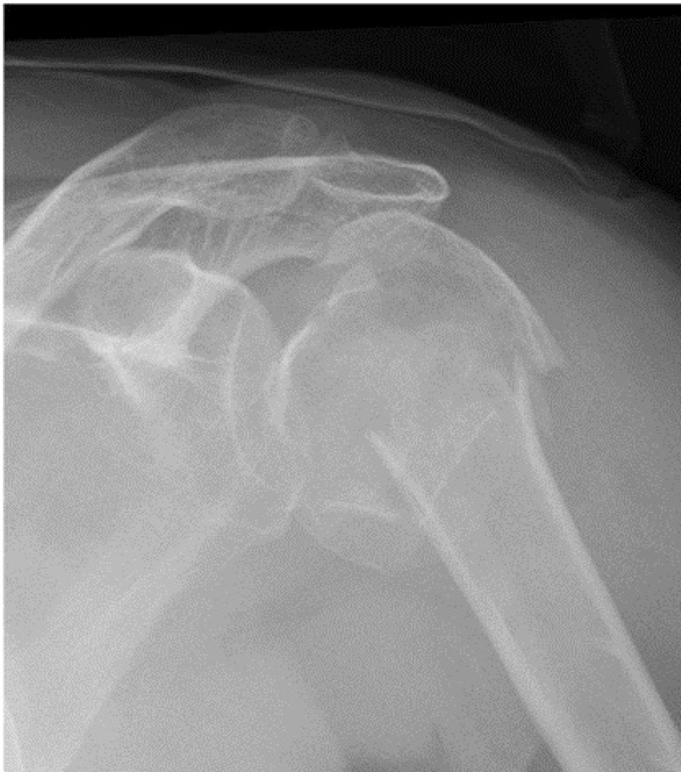
The shoulder is a complex region formed by three bones: the upper end of the arm bone (proximal humerus), the shoulder blade (scapula) and the collar bone (clavicle). Fractures around the shoulder involve the proximal humerus, the clavicle and the scapula (including the glenoid). Proximal humerus fractures are the most common, occurring particularly in women over 60 after low-energy falls related to osteoporosis. Clavicle fractures are common in young, active patients following sports injuries and road traffic accidents. Scapular fractures occur in high-energy injuries and are usually associated with chest and lung injuries. Most fractures around the shoulder are suitable for non-operative treatment; however, surgery is required for displaced, unstable and intra-articular fractures.
Anatomy of the Shoulder
The humeral head sits in the shallow socket of the scapula (the glenoid) to form the ball-and-socket glenohumeral joint. The joint surfaces are covered with cartilage; the labrum deepens the socket, and the rotator cuff muscles (supraspinatus, infraspinatus, teres minor and subscapularis) hold the humeral head within it.
The clavicle connects the chest wall to the shoulder and provides a stable strut for shoulder movement. The scapula glides over the chest wall, enabling the wide arc of shoulder motion. A healthy shoulder allows raising the arm, reaching overhead, throwing and the fundamental movements of daily life without pain.
Proximal Humerus Fractures
These are fractures of the region of the humerus close to the shoulder. They may occur after a fall or a high-energy injury. The Neer classification is based on the number of fracture parts:
- One-part: displacement <1 cm or angulation <45°; may be treated non-operatively in some cases
- Two-part: a single anatomical part is displaced; surgery may be required
- Three-part: two anatomical parts are displaced; surgery is often required
- Four-part: all four anatomical parts are displaced; the blood supply to the humeral head may be compromised, and surgery (plate and screws or arthroplasty) is required
- Fracture–dislocation: fracture plus shoulder dislocation; urgent treatment is required
- Anatomical neck fracture: the blood supply to the humeral head is disrupted; the risk of avascular necrosis (bone death) is high
- Surgical neck fracture: the most common two-part pattern; surgical treatment often gives a good result
- Greater tuberosity fracture: the attachment site of the rotator cuff; displacement >5 mm requires surgery
- Lesser tuberosity fracture: the attachment site of subscapularis; uncommon
Clavicle (Collar Bone) Fractures
Clavicle fractures are divided into three regions according to the Allman / Robinson classification:
- Middle third (mid-shaft): the most common type (~80%); usually treated non-operatively, although surgery is preferred where there is marked displacement, comminution or shortening
- Distal (lateral) third: close to the acromioclavicular joint; surgery is required for displaced type II injuries (with rupture of the coracoclavicular ligaments)
- Medial third: close to the sternum; uncommon, high energy, and requires attention to possible mediastinal injury
Scapula (Shoulder Blade) Fractures
Scapular fractures account for less than 1% of all fractures; they usually result from high-energy injuries and require attention to associated injuries.
- Scapular body fracture: the most common type; most are treated non-operatively
- Glenoid (socket) fracture: intra-articular; surgery is required for displaced fragments, and some patterns may lead to shoulder instability
- Scapular neck fracture: surgery may be required where there is displacement and angulation
- Acromion fracture: may affect the rotator cuff outlet
- Coracoid process fracture: the attachment site of ligaments and tendons; uncommon
- "Floating shoulder": clavicle plus scapular fracture on the same side; more often requires surgery
Classification by Displacement and Joint Involvement
- Non-displaced fracture: the bone fragments have not shifted; most are suitable for non-operative treatment
- Displaced fracture: the fragments have shifted; surgical correction may be required
- Intra-articular fracture: the fracture line extends into the joint surface, increasing the long-term risk of arthritis
- Open fracture: uncommon; requires emergency surgery
- Stress fracture: caused by repetitive loading; may be seen in the clavicle, in athletes and in older patients
Symptoms
- Sudden, severe pain: at the moment of injury, prominent over the shoulder or collar bone
- Swelling and bruising: oedema and haematoma developing within the first hours
- Restricted movement: inability to raise the arm, difficulty with internal and external rotation
- Deformity: a visible prominence in displaced clavicle fractures (a "tenting" appearance); loss of the normal contour around the shoulder in proximal humerus fractures
- Tenderness: pain that increases on palpation of the fracture site
- Crepitus: a grating sensation or sound during movement
- Supporting the arm: the patient holds the arm close to the body with the other hand
- Altered sensation or coldness: if there is nerve or vascular injury (particularly the axillary nerve or brachial plexus)
- Open wound: loss of skin integrity in an open fracture
- Shortness of breath or chest pain: important as an indicator of associated lung or chest wall injury in scapular or high-energy fractures
Mechanisms of Injury
- Low energy: tripping or falling on level ground, falling onto the elbow or an outstretched hand (typical of proximal humerus fractures in older patients with osteoporosis)
- High energy: road traffic accidents (particularly motorcycle and bicycle), falls from height
- Direct blow: a direct impact to the shoulder or collar bone (typical of clavicle and scapular fractures)
- Sports injuries: falls from a bicycle, skiing, motocross, rugby, football
- Pathological fractures: with minimal trauma in the presence of bone tumour or metastasis
- Repetitive loading (for stress fractures)
Diagnosis
Diagnosis is based on a detailed history, physical examination and imaging. Examination assesses swelling, deformity, points of tenderness, range of motion, associated ligament injuries, neurovascular status (particularly the axillary nerve — sensation over the outer upper arm and deltoid contraction), pulses, skin integrity and the chest (for pneumothorax in scapular and high-energy injuries).
Plain radiographs are the first-line investigation; anteroposterior, scapular Y and axillary views for the proximal humerus, anteroposterior and cephalic tilt views for the clavicle, and anteroposterior and axillary views for the scapula are standard. CT is almost always required for intra-articular fractures (particularly glenoid and comminuted proximal humerus fractures) and for surgical planning; three-dimensional CT is critical for assessing fragment displacement. MRI is used to evaluate associated rotator cuff injury and soft-tissue pathology. Angiography or CT angiography may be required where vascular injury is suspected after a high-energy injury.
Non-operative (Conservative) Treatment
A sling or a body–arm bandage (Velpeau bandage) is used for stable, non-displaced or minimally displaced fractures. The sling is worn for an average of 3–6 weeks, after which graduated passive range-of-motion exercises begin.
- Non-displaced proximal humerus fractures (Neer one-part)
- Isolated minimally displaced mid-shaft clavicle fractures
- Most scapular body fractures
- Non-displaced glenoid fractures
Surgical Treatment (ORIF or Arthroplasty)
Surgery is performed for displaced or unstable fractures and for those that disturb the joint surface. Plates and screws, intramedullary nails, tension band techniques, cannulated screws, reverse shoulder arthroplasty or anatomical shoulder arthroplasty may be used.
- Displaced Neer three- and four-part proximal humerus fractures
- Comminuted four-part fractures in older patients (usually with reverse shoulder arthroplasty)
- Displaced mid-shaft clavicle fractures (>2 cm shortening, >100% displacement, comminuted fracture)
- Displaced distal clavicle type II fractures
- Displaced glenoid fractures (particularly of the anterior rim)
- Marked angulation in a scapular neck fracture; open fractures; cases with associated vascular or nerve injury
Advantages
The principal advantage is anatomical restoration of the fracture with rigid fixation. This reduces the long-term risk of malunion, non-union, disturbance of shoulder mechanics and post-traumatic arthritis. Whereas prolonged immobilisation in a sling during non-operative treatment can lead to shoulder stiffness, surgery allows early movement.
- Anatomical reduction: fragments are restored with millimetre precision and the joint surface is accurately repaired
- Early motion: stable fixation allows passive and active shoulder range-of-motion exercises to begin early, reducing stiffness related to sling immobilisation
- Prevention of clavicular shortening: in markedly shortened mid-shaft clavicle fractures, surgery restores anatomical length and improves shoulder mechanics
- Operative time: 60–90 minutes for clavicle fractures; 90–150 minutes for proximal humerus fractures; longer for reverse arthroplasty or complex cases
- Discharge after 1–2 days
- Shoulder function is better preserved in the long term; the risk of non-union and loss of function is higher after non-operative treatment of displaced fractures
Disadvantages
- You will need to wear a sling; moving the arm excessively during this period may lead to implant failure or loss of reduction in the early phase
- There are surgery-specific risks: wound infection, deep infection, implant failure, nerve injury (particularly the axillary nerve), rare vascular injury and rare anaesthetic complications
- Avascular necrosis of the humeral head: the blood supply may be compromised, particularly in anatomical neck and four-part fractures; collapse of the humeral head may occur even after ORIF and conversion to arthroplasty may be required
- Implant irritation: plates over the clavicle may cause irritation in patients with thin skin; implant removal may be required in 20–30% of patients once the bone has healed
- Shoulder stiffness may develop during immobilisation; frozen shoulder (adhesive capsulitis) may occur, particularly in older patients. This resolves with physiotherapy.
Why Surgery?
The shoulder has the widest range of motion of any joint in the body and is critical to many activities of daily life. In proximal humerus fractures, displaced fragments impair rotator cuff function, and advanced displacement threatens the blood supply to the humeral head.
Marked shortening in a clavicle fracture disturbs shoulder mechanics and scapular movement; the non-union rate in markedly displaced mid-shaft clavicle fractures treated non-operatively reaches 15–20%, whereas after surgery it falls below 5%. In glenoid fractures, incongruity of the joint surface leads to shoulder arthritis in the long term.
Who is a Candidate?
Surgery is performed for displaced or unstable fractures around the shoulder and for those that disturb the joint surface.
- Displaced Neer three- and four-part proximal humerus fractures
- Greater tuberosity fractures displaced >5 mm
- Markedly shortened (>2 cm) or comminuted mid-shaft clavicle fractures
- Displaced distal clavicle type II fractures
- Glenoid fractures involving the joint surface; open fractures
- Complex cases such as neurovascular injury or a "floating shoulder"
- Non-operative treatment: stable, non-displaced fractures; selected patients at very high surgical risk; most scapular body fractures
Expected Outcomes
Most patients with a correctly treated fracture around the shoulder achieve a pain-free, functional shoulder. Most return to daily activities within 3–6 months; complete recovery may take up to a year in some cases. Fracture healing takes 6–8 weeks in simple isolated fractures and 10–12 weeks or longer in complex or intra-articular fractures.
Success rates after surgery for clavicle fractures are high; in comminuted proximal humerus fractures in older patients, reverse shoulder arthroplasty has in recent years become an option that gives very satisfactory results.
Surgery & First 3 Weeks
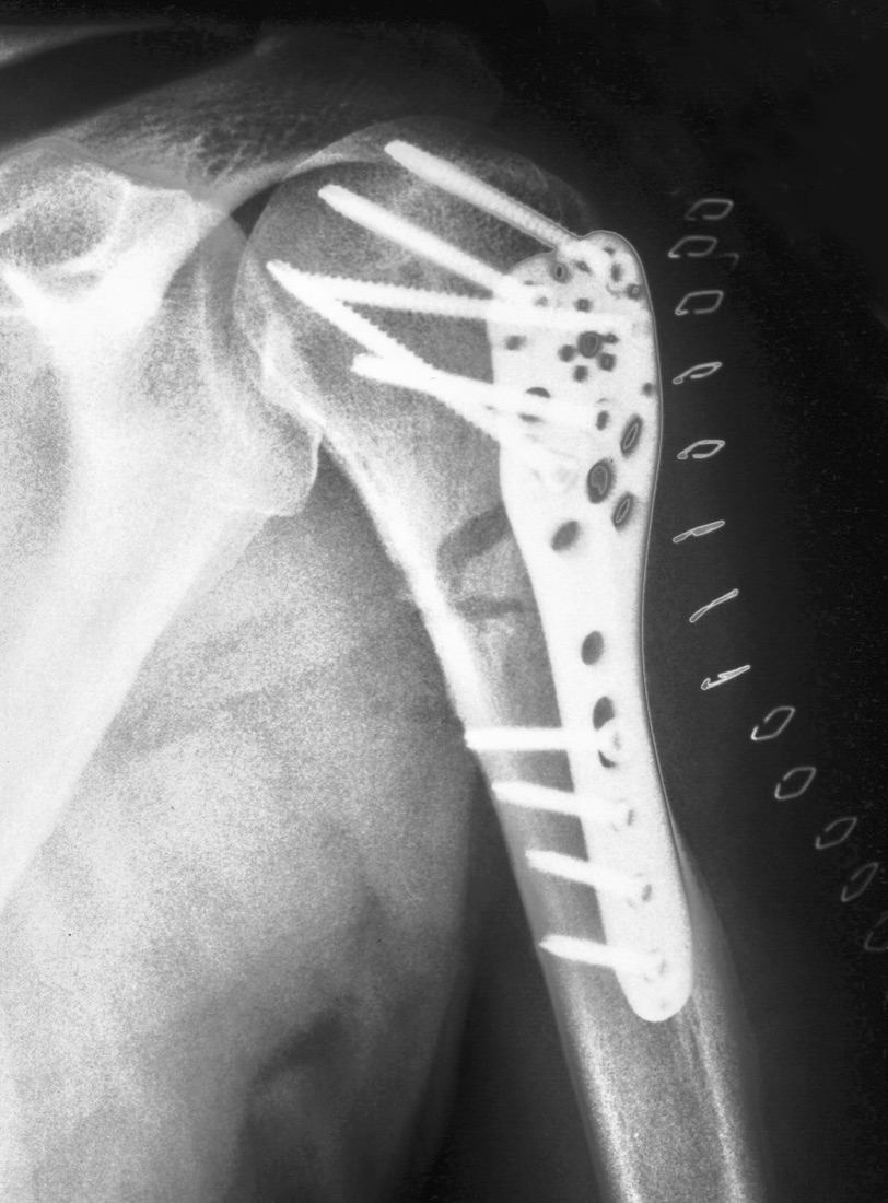
There are two main approaches to the treatment of fractures around the shoulder: non-operative (sling or bandage) and surgical (open reduction and internal fixation — ORIF, or arthroplasty). The choice depends on the fracture type, its stability and patient factors.
Non-operative Treatment
A sling or a body–arm bandage (Velpeau bandage) is used for stable, non-displaced fractures.
- Assessment in the emergency department; reduction under anaesthesia if required
- A sling or Velpeau bandage is applied
- Sling use for the first 3–6 weeks; graduated passive range-of-motion exercises as permitted by the surgeon
- Ice and analgesia
- Follow-up radiographs at weeks 1, 2, 4, 6 and 8 to monitor for displacement
- Graduated active movement and physiotherapy once the sling is discontinued
Surgical Treatment — General Information
Surgery is performed under general anaesthesia, frequently combined with an interscalene block. The block is effective for pain control in the first 24–48 hours. Radiographs and CT are reviewed for preoperative planning; three-dimensional CT is used for detailed planning in comminuted proximal humerus and glenoid fractures. Prophylactic antibiotics are given.
- Duration: 60–90 minutes for clavicle fractures; 90–120 minutes for proximal humerus fractures; 120 minutes or more for reverse shoulder arthroplasty and complex cases
- Anaesthesia: general anaesthesia plus interscalene block
- Hospital stay: discharge the following day (for clavicle and selected proximal humerus fractures) or 2 days; longer in complex and arthroplasty cases
Surgical Approaches (Incisions)
- Deltopectoral approach: the most widely used approach for proximal humerus fractures and shoulder arthroplasty; a 10–15 cm incision at the front of the shoulder
- Anterolateral (deltoid-splitting) approach: for greater tuberosity fractures and some proximal humerus fractures; an incision of approximately 5 cm on the outer side of the shoulder
- Longitudinal incision over the clavicle: for clavicle fractures
- Posterior (Judet) approach: for scapular and glenoid fractures
- Minimally invasive percutaneous techniques: K-wires or screws placed through small incisions in selected proximal humerus fractures
Proximal Humerus Fracture — Technique
- The patient is positioned in the beach-chair position and a deltopectoral incision is made
- The fracture fragments are reduced anatomically and the rotator cuff attachment sites (greater and lesser tuberosities) are restored to their anatomical positions
- Fixation is achieved with a lateral locking plate (a proximal humeral locking plate such as PHILOS) and multidirectional locking screws
- Alternative: an intramedullary nail (particularly in extra-articular two-part fractures)
- Percutaneous (closed) technique: fixation with K-wires or cannulated screws, usually in adolescent or paediatric patients with minimal comminution
Hemiarthroplasty or Reverse Shoulder Arthroplasty
- Arthroplasty may be preferred in four-part fractures, anatomical neck fractures (with a high risk of avascular necrosis) and comminuted fractures in older patients
- Anatomical / hemiarthroplasty: replacement of the humeral head with a prosthesis; may be considered where the rotator cuff is intact
- Reverse shoulder arthroplasty (RSA): the preferred option in recent years, giving good results particularly in older patients and where rotator cuff quality is uncertain
Clavicle Fracture — Technique
- The patient is positioned supine with the head turned away; a longitudinal incision is made over the clavicle
- The fracture fragments are reduced anatomically
- Fixation is achieved with a superior or anterior locking plate and screws
- Alternative: an elastic intramedullary nail (particularly in children and young athletes)
- For distal clavicle type II fractures, dedicated distal clavicle plates or coracoclavicular fixation techniques (TightRope, coracoid sling) are used
Scapula / Glenoid Fracture — Technique
- The patient is positioned in the lateral decubitus position; a posterior (Judet) approach is used, or a deltopectoral approach for the anterior glenoid rim
- In glenoid fractures, displaced fragments are fixed with cannulated screws or mini-plates
- In scapular neck fractures, anatomical reduction is achieved with plates and screws
Open Fracture
- Emergency debridement and irrigation, with antibiotic and tetanus prophylaxis
- Depending on the soft-tissue condition, either early definitive fixation or a staged approach with temporary external fixation
Implant Options
- Plates: proximal humeral locking plates (PHILOS type); clavicle plates (superior or anterior, anatomically contoured, locking); distal clavicle plates (hook plate); mini-plates for the scapula and glenoid
- Screws: cortical screws (3.5 / 4.5 mm); cannulated screws (greater and lesser tuberosities, glenoid fragments); locking screws
- K-wires (for percutaneous technique and tension band)
- Intramedullary nail (for clavicle and proximal humerus fractures)
- TightRope / coracoid sling systems (for distal clavicle fractures)
- Reverse shoulder arthroplasty / hemiarthroplasty (for complex proximal humerus fractures)
- External fixator (rarely, in open fractures)
Discharge Information
Patients are usually discharged the following day (for clavicle and selected proximal humerus fractures) or after two days; the stay may be longer after reverse shoulder arthroplasty or in complex cases. After surgery the shoulder is supported in a sling, with an abduction pillow added at the surgeon's discretion.
Before discharge, the physiotherapist teaches how to apply and remove the sling, pendulum (Codman) exercises, elbow, wrist and finger range-of-motion exercises and daily transfers (bathing, dressing). Sling use is obligatory for the first 4–6 weeks after surgery and active use of the arm is prohibited; only passive range-of-motion exercises are started, with the surgeon's permission.
Ice for 15–20 minutes five times a day is recommended after discharge. Analgesics, anti-inflammatories and gastric protection are prescribed. Dressings are changed regularly; it is important not to wet the wound for the first 10–14 days, and sutures are removed on day 14. Sleeping semi-upright (supported with pillows) is recommended.
Early Postoperative Period (First 3 Weeks)
The aims during this period are wound healing, control of swelling, pain management, preserving elbow, wrist and finger movement, minimising muscle atrophy and supporting healing of the soft tissues around the shoulder. Weekly review is recommended.
- Wound monitoring: the skin over the clavicle and shoulder is relatively thin. Report discharge, marked redness, increased warmth, wound edge separation or malodour immediately
- Sling use: the sling is usually worn full time for 4–6 weeks, including at night. Active elevation of the arm is prohibited for the first 6 weeks
- Passive movement: with the surgeon's permission, passive pendulum (Codman) and gentle flexion exercises may begin in the first weeks. Active movement and resisted work are prohibited
- Elbow, wrist and finger movement: performed three to four times a day within the sling; this reduces swelling and prevents muscle atrophy
- Pain: once the interscalene block wears off, prescribed medication, ice and an appropriate resting position control the pain
- Nerve and vascular monitoring: the axillary nerve is the nerve most commonly affected in shoulder surgery; sensation over the outer upper arm and deltoid contraction are checked. Sensation and strength in the hand and fingers are assessed for the brachial plexus, and the radial pulse and hand colour are monitored
Follow-up
- Days 10–14: suture removal and radiographic review
- Weeks 4–6: decision on discontinuing the sling
- Month 3: assessment of bone healing
- Month 6 and annually thereafter if required
Risks & Complications
The treatment of fractures around the shoulder is well standardised in modern orthopaedics and most patients do well. Nevertheless, both sling treatment and surgery carry specific risks, and it is important that you understand the possible complications.
Risks Specific to Non-operative (Sling) Treatment
- Inadequate reduction / secondary displacement: fragments shifting within the sling; further surgery may be required
- Skin problems: pressure sores and friction beneath the sling
- Shoulder stiffness / frozen shoulder (adhesive capsulitis): the most common complication, particularly in older patients
- Rotator cuff weakness: atrophy related to immobilisation
- Non-union: may reach 15–20%, particularly in displaced mid-shaft clavicle fractures
- Malunion: shortening and deformity of the clavicle with disturbance of shoulder mechanics
Risks Specific to Surgery — Common (Under 5%)
- Superficial wound infection (usually treated with antibiotics)
- Postoperative pain, swelling and temporary bruising
- Temporary joint stiffness — resolves with regular physiotherapy
- Temporary loss of strength — regained with rehabilitation
- Transient symptoms related to anaesthesia such as nausea and dizziness
- Temporary altered sensation after an interscalene block
- Scar discomfort (particularly over the clavicle)
Risks Specific to Surgery — Less Common
- Deep infection (osteomyelitis / implant infection): 1–3%; may require revision, implant removal and prolonged antibiotics
- Nerve injury: the axillary nerve (the most commonly affected in shoulder surgery), the suprascapular nerve and elements of the brachial plexus
- Vascular injury (very rare, under 1%)
- Pneumothorax (particularly with clavicle and scapular fractures)
- Implant failure: loosening or breakage of screws or plates, particularly in osteoporotic bone
- Avascular necrosis (bone death of the humeral head): particularly in anatomical neck and four-part fractures; may develop even after ORIF, with rates reaching 25–30%
- Heterotopic ossification (bone formation in soft tissue)
- Complex regional pain syndrome (CRPS / Sudeck's atrophy): skin colour change, pain and temporary bone loss. This is a fully reversible condition
- Deep vein thrombosis (DVT) / pulmonary embolism — rare in upper limb surgery
Long-term Complications
- Malunion: the bone heals in the wrong position, disturbing shoulder mechanics and increasing pain and the risk of arthritis; this is common with clavicular shortening
- Non-union: increased by smoking, diabetes, osteoporosis and early activity
- Post-traumatic osteoarthritis: may develop years later after intra-articular fractures (particularly glenoid and humeral head fractures); shoulder arthroplasty may be required in advanced cases
- Persistent shoulder stiffness (frozen shoulder)
- Rotator cuff weakness or tearing
- Chronic pain; chronic instability (particularly after glenoid fractures)
- Cosmetic deformity (in clavicle fractures)
Implant-related Problems
- Implant irritation close to the skin: a plate over the clavicle causes marked irritation in patients with thin skin; implant removal may be required in 20–30% of patients
- Need for implant removal: usually performed after 9–12 months, once fracture healing is complete
- Screw cut-out (a screw penetrating into the joint in an osteoporotic humerus): may occur with proximal humeral locking plate fixation
Factors that Increase Risk
- Smoking (markedly increases the risk of wound healing problems, non-union and infection)
- Poorly controlled diabetes (impaired wound healing)
- Osteoporosis (poor implant purchase, secondary displacement, screw cut-out)
- Peripheral vascular disease
- Immunosuppressive therapy and rheumatological disease
- Advanced age; high-energy or open fracture
- Active or recently treated infection (dental, urinary, skin)
Contact Your Doctor Immediately If You Have
- Sudden increase in pain or swelling
- Fever above 38 °C
- Discharge, malodour or separation of the wound
- Numbness, tingling or loss of strength in the arm, shoulder or hand
- Bruising, coldness or pallor of the hands or fingers (suspected vascular problem)
- Shortness of breath or chest pain (suspected pneumothorax or pulmonary embolism)
- Wound edge separation or exposed hardware
- Sudden change in the shape of the shoulder
Long-term Outcomes
With accurate diagnosis, appropriate treatment and compliant rehabilitation, the great majority of fractures around the shoulder heal without problems. Most patients return to daily activities within 3–6 months and to sport within 6–12 months. Complete recovery may take up to a year in some cases; stiffness may persist for 12 months, particularly after high-energy and complex proximal humerus fractures.
There is a risk of post-traumatic arthritis years later after intra-articular fractures; anatomical reduction, appropriate implant selection, early motion and compliant rehabilitation reduce this risk. Stopping smoking, controlling blood glucose and maintaining a healthy weight are critical factors for healing and long-term outcome.
Rehabilitation
Rehabilitation after a fracture around the shoulder is the most important part of recovery. The aims are to control pain, progressively restore shoulder range of motion, strengthen the rotator cuff and scapular stabilisers, and return the patient to daily life and sporting activity. Because shoulder stiffness (frozen shoulder) is the most common complication after these fractures, beginning passive range-of-motion exercises as soon as it is safe to do so is critical.
Rehabilitation After Non-operative Treatment
- Weeks 0–2: Sling. Ice and pain control. Active elbow, wrist and finger range-of-motion exercises. Pendulum (Codman) exercises may begin with the surgeon's permission.
- Weeks 2–6: The sling is discontinued gradually (according to fracture type). Passive pendulum and gentle flexion and external rotation exercises within the limits set by the surgeon. Active movement is prohibited.
- Weeks 6–12: The sling is fully discontinued. Active-assisted and active range-of-motion exercises begin. Light rotator cuff and scapular stabilisation exercises.
- Months 3–4: Full active range of motion is the target. Graduated strengthening (resistance band, light weights).
- Months 4–6: Proprioception and dynamic stabilisation training. Introduction of sport-specific drills.
- Months 6–12: Return to light recreational sport; full return to contact and throwing sport with the surgeon's approval.
Rehabilitation After Surgery (ORIF)
- Weeks 0–2: Sling full time. Ice and pain control. Active elbow, wrist and finger range-of-motion exercises. With the surgeon's permission, passive pendulum (Codman) and gentle flexion exercises may begin. Suture removal at days 10–14.
- Weeks 2–6: The sling is discontinued gradually (according to fracture type). Passive range-of-motion exercises are increased progressively (flexion, abduction and external rotation within the limits set by the surgeon). Scapular isometric exercises.
- Weeks 6–12: The sling is fully discontinued. Active-assisted and active range-of-motion exercises begin; full range of motion is the target. Light rotator cuff and scapular stabilisation exercises (resistance band).
- Months 3–4: Full active range of motion is maintained. Higher-resistance strengthening. Swimming (progressively) and cycling are recommended. Weight lifting is prohibited.
- Months 4–6: Proprioception and dynamic stabilisation training. Introduction of plyometric exercises.
- Months 6–9: Sport-specific drills; throwing mechanics and overhead movements are introduced gradually. Return to low-contact sport.
- Months 9–12: Full return to contact, throwing and overhead sport with the surgeon's approval and once functional criteria are met.
Rehabilitation After a Clavicle Fracture (Special Notes)
- The sling is worn for 3–6 weeks (longer with non-operative treatment, and possibly shorter after surgery)
- Pendulum exercises may begin from the first week
- Active range of motion is usually fully restored by weeks 6–8
- Shoulder stiffness is less common after a clavicle fracture than after a proximal humerus fracture, and recovery is generally faster
Criteria for Return to Sport and Activity
- Full, pain-free shoulder range of motion (flexion ≥160°, abduction ≥150°, external rotation ≥60°)
- Rotator cuff and scapular muscle strength at least 85–90% of the opposite shoulder
- No swelling or pain after activity
- Pain-free completion of sport-specific drills
- Control of throwing mechanics
- Approval of the surgeon and physiotherapist
Recommended Activities
- Stationary cycling, elliptical trainer, walking
- Swimming (once the wound has healed; progressively)
- Low-impact conditioning and strengthening exercise
- Pilates and yoga (with the surgeon's permission)
- Walking and light jogging (from month 3–4)
Activities to Avoid in the First 3–6 Months
- Lifting weights overhead
- Sudden pulling or tugging movements
- Contact sport (rugby, handball, basketball)
- Throwing sports (baseball, volleyball, handball)
- Lying on the operated shoulder
- Cycling (risk of falling, particularly with clavicle fractures)
Lifestyle and Recovery Advice
- Perform your home exercise programme every day — it is the single most important determinant of long-term outcome
- Do not smoke (markedly increases the risk of non-union, wound healing problems and infection)
- Maintain tight blood glucose control if you have diabetes
- Healthy weight: important for overall recovery
- Sleeping semi-upright (supported with pillows) is recommended in the first weeks
- Avoid lying on the operated shoulder for the first 3 months; lie on your back or on the opposite side
- Attend your follow-up appointments (weeks 4–6, month 3, month 6, year 1)
Expected Outcomes
Most patients return to pain-free daily activity 3–6 months after treatment of a fracture around the shoulder. Mild stiffness and a small loss of range of motion may persist for 6–12 months; this is expected and treatable.
Return to sport and high-demand activity occurs between months 6 and 12; recovery may take longer after high-energy or comminuted fractures. Patient satisfaction is high with modern treatment methods and disciplined rehabilitation; anatomical reduction, early passive motion and appropriate rehabilitation reduce the long-term risk of stiffness and loss of function.
Is This Treatment Right for You?
Every patient's condition is different. After a detailed examination and imaging, the treatment plan that suits you best will be prepared.
Would you like more information about Proximal Humerus Fracture?
Book Appointment