Overview
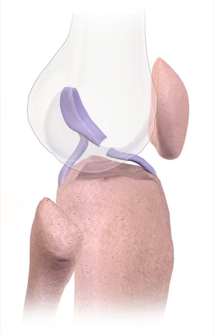
The anterior cruciate ligament (ACL) is one of the four main ligaments of the knee, running diagonally in the centre of the joint between the thigh bone (femur) and the shin bone (tibia). It is approximately 3 cm long, thick and taut. The ACL is the principal stabiliser of the knee, preventing the tibia from sliding forwards on the femur and limiting excessive rotation. The great majority of ACL injuries are complete or near-complete tears; partial tears are uncommon.
Function of the ACL
- Anteroposterior stability: prevents the tibia from sliding forwards on the femur. The ACL resists approximately 85% of the total anterior translational force.
- Rotational stability: limits excessive internal and external rotation with the knee flexed, and controls the knee during pivoting movements.
- Load distribution: works together with the menisci to contribute to balanced load transfer across the knee.
- Proprioception: nerve endings within the ligament inform the central nervous system of the position of the knee in space; after a tear this sense is disturbed and a feeling of instability develops.
- Healing potential: because the ACL is an intra-articular ligament bathed in joint fluid, it does not heal spontaneously once torn. In active patients the treatment is therefore usually not to suture the ligament but to reconstruct it using another tendon (a graft).
Types of Tear
- Complete tear: the ligament is completely ruptured; this is the most common pattern and reconstruction is recommended
- Partial tear: some ligament fibres remain intact; this is uncommon and non-operative management may be appropriate in some cases
- Tibial avulsion: the ligament pulls away with a small fragment of bone on the tibial side; seen particularly in children, where primary repair may be appropriate
- Femoral avulsion: the ligament pulls away from the femoral side; rare
Symptoms and How the Diagnosis is Made
Symptoms vary according to whether the injury is acute or chronic and whether there is associated damage.
- A "pop": a sensation or sound of rupture from within the knee at the moment of injury
- Rapid swelling: marked haemarthrosis (bleeding into the joint) within the first 6–24 hours
- Inability to bear weight: unable to take weight on the leg; a feeling that the knee will not hold
- Restricted movement: difficulty fully straightening or bending the knee because of swelling
- Instability or giving way: particularly when pivoting, squatting or descending stairs
- Joint line pain: prominent where there is an associated meniscal tear
An Acute Knee Injury is an Emergency
If you experience a pop while pivoting, followed by rapid swelling and an inability to bear weight, you should be assessed as soon as possible; bleeding into the joint, an associated meniscal tear and cartilage damage can be managed with early diagnosis.
Diagnosis
Examination includes the Lachman test (the most sensitive), the anterior drawer test and the pivot shift test; joint effusion and meniscal tenderness are assessed. Plain radiographs show the bone architecture, any associated fractures (particularly a Segond fracture) and the joint space, but do not show the ligament itself. MRI is the gold standard for demonstrating the site and type of tear and any associated meniscal, cartilage or ligament damage. The patient's age, activity level and mechanism of injury (usually a sudden twist or change of direction with the foot planted) are central to the decision. The definitive decision and treatment plan are made at arthroscopy, with the tear seen directly.
Advantages
- Most importantly, reconstruction prevents meniscal and cartilage damage in the long term. In untreated ACL tears, repeated episodes of giving way lead to meniscal tears and early post-traumatic osteoarthritis.
- Operative time: on average 60–120 minutes; general or spinal anaesthesia
- Discharge on the first or second postoperative day
- Because it is performed arthroscopically through small incisions, there is less pain, a lower infection risk and faster early recovery
- Success rate of 85–90% in active patients; return to the previous level of activity is achievable
Disadvantages
- It requires a long and disciplined rehabilitation; return to sport takes on average 9–12 months.
- If an autograft is used, there may be temporary pain, weakness or altered sensation at the donor site (front of the knee or hamstrings).
- Progression to active strengthening is undertaken cautiously until the graft has biologically ligamentised (approximately 6–12 months); re-injury during this period can lead to graft failure.
- Re-rupture occurs in 5–10% of young athletes; smoking, poor rehabilitation and early return to sport increase the risk.
Why ACL Reconstruction?
The ACL has critical roles in knee stability, proprioception and protection of the menisci and cartilage. It does not heal spontaneously once torn. In active individuals, repeated episodes of instability lead to meniscal tears and articular cartilage damage, and to post-traumatic osteoarthritis in the long term. Current AAOS clinical guidance recommends reconstruction within the first 1–3 months after injury in the great majority of young and active patients; this window is critical for preventing meniscal and cartilage damage. In suitable candidates the torn ACL is therefore reconstructed with a graft.
Who is a Candidate?
Arthroscopic ACL reconstruction is performed in patients with a complete — or a functionally inadequate partial — tear who have an active lifestyle.
- Athletes involved in pivoting sports (football, basketball, volleyball, skiing, handball)
- Patients who experience knee instability in daily life or at work
- Those with an associated meniscal tear or cartilage damage
- Patients with recurrent episodes of giving way despite non-operative treatment
- In children and adolescents, using modified techniques (such as over-the-top) that protect the growth plate
Reconstruction with a Graft
The torn ligament is removed; tunnels are created in the tibia and femur and a tendon graft taken either from the patient's own body (autograft) or from a donor (allograft) is positioned as the new ACL and secured with screws or fixation devices.
Surgery & First 3 Weeks
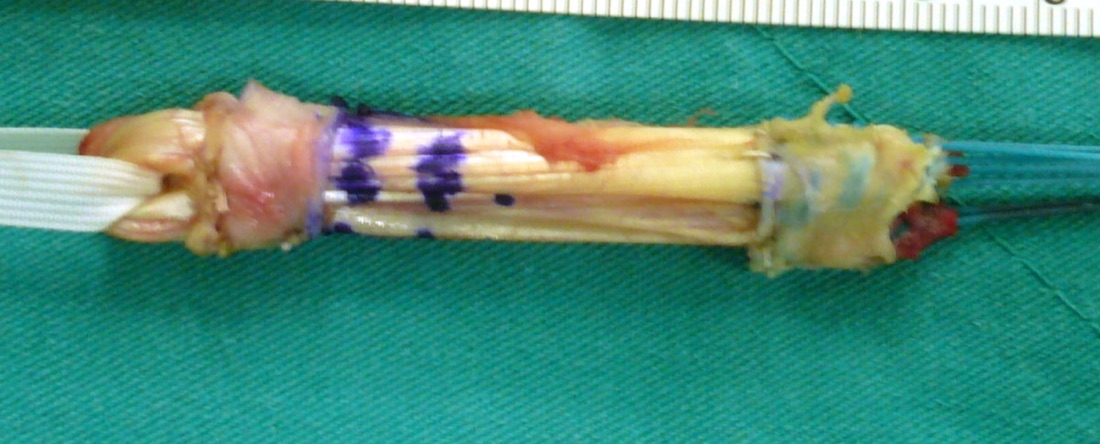
Arthroscopic ACL reconstruction is a procedure in which the torn anterior cruciate ligament is removed and replaced with a tendon graft taken from the patient's own body or from a donor. Rather than being sutured directly, the new graft is passed through precisely drilled bone tunnels in the tibia and femur and secured with bio-absorbable or titanium fixation devices. The knee is entered arthroscopically through small incisions (approximately 1 cm portals); any associated meniscal and cartilage damage is addressed at the same time.
General Information About the Operation
- Aim: to restore knee stability, enable return to activity, and protect the menisci and cartilage from long-term damage
- Duration: 60–120 minutes, depending on any concomitant procedures (meniscal repair, cartilage work)
- Anaesthesia: general or spinal
- Hospital stay: usually day case or discharge the following day
- Timing: recommended at the earliest opportunity once swelling has settled and range of motion has recovered; delay increases the risk of meniscal and cartilage damage
- Complications: the rate of serious complications is below 2%
Graft Selection
Graft choice is individualised according to the patient's age, activity level, occupation, sport, other pathology within the knee, previous surgery and the surgeon's experience. In general, autografts are preferred in young, active patients and allografts in older, lower-demand patients.
Autograft (the Patient's Own Tissue)
- Hamstring (semitendinosus ± gracilis) tendon: the most commonly used autograft. Less anterior knee pain and difficulty kneeling; a small incision is required. In young athletes with high demands, the risk of re-rupture may be slightly higher than with patellar tendon.
- Patellar tendon (bone–patellar tendon–bone / BTB): regarded as the "gold standard". Bone-to-bone healing gives secure fixation, with low rates of re-rupture and infection. Anterior knee pain and difficulty kneeling are more common.
- Quadriceps tendon: an option that has become popular in recent years. It can be harvested with a bone block or as tendon alone. Some studies report a lower re-rupture rate than hamstring graft in young and adolescent athletes, with less anterior knee pain than patellar tendon.
- Iliotibial band (ITB): may be preferred in children and adolescents, using growth-plate-sparing techniques (for example all-epiphyseal reconstruction).
Allograft (Donor Tissue)
- Patellar tendon, Achilles tendon, semitendinosus, gracilis or posterior tibialis allografts may be used; because no harvest incision is needed, operative time and donor-site pain are reduced
- Advantages: smaller incision, less early pain and faster early recovery
- Disadvantages: graft incorporation is slower. The risk of re-rupture is markedly higher than with autograft in young, high-demand athletes, so allograft is not preferred in patients under 25 or in active athletes
- With modern tissue bank processing, the risk of disease transmission is extremely low
Discharge Information
Patients are usually discharged the same day or after one night in hospital. On the day of or the day after surgery you are mobilised with the physiotherapist and begin walking with support. The movement and loading permitted on the operated knee will be explained by your physiotherapist and surgeon according to any additional procedures performed. A brace will be fitted where protection is required.
On the first day the physiotherapist teaches quadriceps isometric exercises, straight leg raises and ankle pumping; it is important to perform these 3–4 times a day.
Cold therapy for 15 minutes over a towel five times a day is recommended after discharge. The medication prescribed includes an anticoagulant, which must be taken for 3 weeks. Anti-embolism stockings must be worn on the operated leg for 3 weeks and on the other leg for 1 week.
Return to Activity
- Strengthening and range-of-motion exercises begin from the first week within a pain-free range
- Quadriceps and hamstring strengthening intensifies in weeks 2–6
- Return to desk-based work usually at weeks 2–3
- Work requiring active use of the knee: weeks 6–8
- Straight-line jogging: month 3
- Graduated return to sport: months 4–6
- Full return to contact and pivoting sport: months 9–12
- Physical activity should be maintained long term and the muscles around the knee kept strong. Weight loss is recommended for patients with a high body mass index.
Early Postoperative Period (First 3 Weeks)
On the day of or the day after surgery you are mobilised with the physiotherapist wearing a brace and begin walking with crutches. Partial weight bearing on the operated leg is permitted as pain allows; after isolated ACL reconstruction most patients can discard the crutches early.
If a meniscal repair was also performed, weight bearing and range of motion are more restricted. Your physiotherapist will show you quadriceps isometric exercises, ankle pumping, straight leg raises and passive/active-assisted knee range-of-motion exercises; these should be performed 4–5 times a day. Regular review is recommended. It is important to wear your compression stockings and take your anticoagulant during the first 3 weeks.
- Wound monitoring: discharge, marked redness and increased warmth are monitored for signs of infection
- Range of motion: full extension is the first goal; 90° of flexion is targeted by week 2. Where a meniscal repair has been performed, flexion is limited to 0–30° for the first 4 weeks and a brace is worn for 6 weeks
- Quadriceps activation: quadriceps inhibition is the single greatest obstacle to recovery. Isometric contractions and straight leg raises should be repeated several times a day
- Pain: unless soft-tissue swelling develops around the knee, there is a marked reduction within 1–2 weeks
- Deep vein thrombosis: clot formation in the deep veins of the leg, diagnosed by Doppler ultrasound. It is very rare and is prevented by compression stockings and anticoagulation
Risks & Complications
Arthroscopic ACL reconstruction is generally a safe and effective procedure. In modern knee surgery the rate of serious complications is below 5%.
Common (Under 5%)
- Superficial wound infection (usually treated with antibiotics)
- Postoperative pain, swelling and temporary bruising
- Quadriceps inhibition / temporary weakness — resolves with physiotherapy
- Joint stiffness and loss of range of motion — largely prevented by intensive physiotherapy
- Donor-site symptoms (with autograft): anterior knee pain (patellar tendon), hamstring weakness, altered sensation
Less Common
- Graft failure / re-rupture: approximately 10% within 10 years; up to 20% in young athletes and as high as 35% in high-demand adolescents
- The risk of re-rupture with allograft is markedly higher in young and active patients than with autograft
- Loosening or migration of a fixation device (screw or button)
- Tunnel widening (particularly important in revision surgery)
- Patellar fracture at the site of the bone block with a patellar tendon graft (very rare)
- Nerve or vascular injury (particularly altered sensation in the saphenous nerve territory; very rare, <1%)
- Deep vein thrombosis / pulmonary embolism (rare)
- Septic arthritis (intra-articular infection) — very rare; a minimally increased risk with allograft
- Growth plate injury in paediatric patients (minimised with appropriate technique)
- Risk of post-traumatic osteoarthritis in the long term (reduced by reconstruction but not eliminated)
Contact Your Doctor Immediately If You Have
- Sudden increase in pain or swelling
- Fever above 38 °C
- Firm, one-sided swelling or redness of the leg, or shortness of breath
- Discharge or malodour from the wound
- An episode of giving way, or another "pop" in the knee
- Numbness, tingling or loss of strength in the leg
Rehabilitation
Rehabilitation after ACL reconstruction is a long and disciplined process and is the single most important determinant of outcome. The programme is individualised according to graft type, any associated meniscal repair and the patient's goals.
Rehabilitation Timeline
- Weeks 0–2: Ice, elevation, compression. A brace is fitted if a meniscal repair was also performed. Full extension is the goal. Quadriceps isometric exercises, ankle pumping, straight leg raises. If no meniscal repair was performed, partial weight bearing with crutches as pain allows; if a meniscal repair was performed, no weight bearing for the first 6 weeks.
- Weeks 2–6: Range of motion is opened progressively to 0–120°. Physiotherapy begins. Progression to full weight bearing (where no meniscal repair was performed). Stationary cycling begins. Quadriceps and hamstring strengthening; hip and core stability work.
- Weeks 6–12: Full range of motion. The brace, if used, is discontinued. Closed-chain exercises (mini squats, leg press) and swimming are recommended. Straight-line treadmill work. Twisting and pivoting are prohibited.
- Months 3–5: Straight-line jogging begins. Higher-resistance strengthening; single-leg exercises. Proprioception and balance training.
- Months 5–8: Pivoting and cutting drills are introduced progressively. Plyometric exercises. Sport-specific open-field work.
- Months 9–12: Full return to contact and pivoting sport, once return-to-sport testing (strength, hop tests, functional tests) is complete and with the surgeon's approval.
Criteria for Return to Sport
- Quadriceps strength at 90% of the opposite leg (LSI ≥ 90%)
- An adequate hamstring-to-quadriceps ratio
- Full, pain-free range of motion
- 90% symmetry on single-leg hop tests (single hop, triple hop, crossover hop, timed hop)
- No swelling or pain after activity
- Control of pivoting, cutting and landing technique; psychological readiness
Knee Health After Reconstruction
A successful ACL reconstruction preserves knee stability and protects the menisci and cartilage from long-term damage. The following measures reduce the risk of re-rupture and post-traumatic osteoarthritis:
- Healthy weight: every additional kilogram places four times that load on the knee
- Maintaining quadriceps, hamstring, hip and core strengthening
- Low-impact sports: swimming, cycling, elliptical trainer
- Avoid high-impact and pivoting sports for the first 9–12 months
- The risk of injury to the opposite knee is also high; strengthen both sides equally
- Stopping smoking (it adversely affects graft integration)
Lifestyle Advice
- Warm up before exercise and stretch afterwards
- Avoid squatting and kneeling for the first 4 months
- Descend stairs slowly and under control
- Follow injury-prevention training programmes (cutting and landing technique) when returning to sport
- Attend your follow-up appointments
Is This Treatment Right for You?
Every patient's condition is different. After a detailed examination and imaging, the treatment plan that suits you best will be prepared.
Would you like more information about ACL Reconstruction?
Book Appointment