Overview
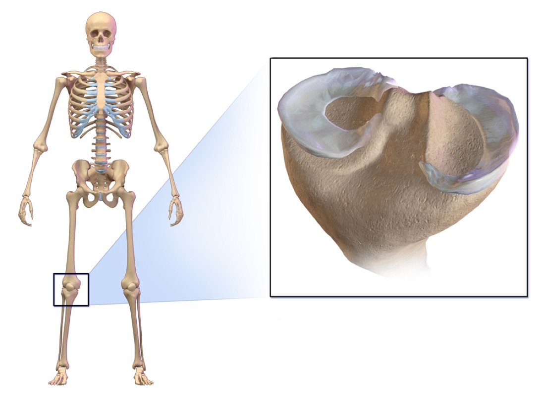
The two crescent-shaped menisci (medial/inner and lateral/outer) are flexible fibrocartilage structures that cover approximately 70% of the opposing surfaces of the tibia and femur. About 50–70% of the load transmitted through the knee passes through the menisci. Any reduction in meniscal surface area (for example after partial meniscectomy) increases the load carried by the articular cartilage.
Functions
- Shock absorption; limiting the end range of flexion and extension
- Load transfer and reduction of the stress transmitted across the joint
- Stabilisation of the knee joint, nutrition of the cartilage and lubrication of the joint surfaces
Healing Potential
The meniscus is divided into three zones according to blood supply:
- Red–red zone (peripheral 10–30%): fully vascularised — high repair success rate
- Red–white zone (border region): partially vascularised — limited healing after repair
- White–white zone (central): avascular — healing after repair is less likely
Types of Tear
- Bucket-handle tear: causes locking of the knee; typically acute and seen in young athletes
- Radial (vertical) tear: functionally divides the meniscus and disrupts load transmission
- Horizontal tear: degenerative, seen in middle-aged and older patients
- Complex / degenerative tear: tearing in more than one plane; seen over the age of 40 and associated with osteoarthritis
Symptoms and How the Diagnosis is Made
Symptoms vary with the size, type and location of the tear and with patient factors such as age and weight.
- Joint line pain: on the inner or outer side of the knee, particularly with bending and twisting
- Swelling: oedema developing within 24–72 hours
- Restricted movement: difficulty achieving full flexion or full extension
- Locking: a bucket-handle tear may cause sudden, complete locking of the knee
- Clicking or catching: a mechanical sensation and sound during movement
- Instability: a feeling of giving way when descending stairs, squatting or pivoting
Locking is an Emergency
If the knee locks completely, seek assessment as soon as possible; a displaced bucket-handle tear may require urgent treatment.
Diagnosis
Examination includes the McMurray and Apley tests. Tenderness along the joint line is an important finding. Plain radiographs are obtained to assess the bone and joint space. MRI is the gold standard for determining the type and size of the tear and whether it is amenable to repair. The definitive decision and treatment plan are made at arthroscopy, with the tear seen directly.
Treatment Options
Arthroscopic meniscal repair is performed for tears located in the red–red or red–white zone of the meniscus — that is, where the blood supply is sufficient for the tissue to be prepared, sutured and expected to heal.
Partial meniscectomy is performed for tears in the red–white or white zone, where the blood supply is insufficient and spontaneous healing cannot be expected.
Advantages — Meniscal Repair
- The most important advantage is prevention of cartilage damage in the long term. Without surgery the knee develops osteoarthritis more quickly.
- Operative time: on average 30–60 minutes; general or spinal anaesthesia
- Discharge the following day
Advantages — Partial Meniscectomy
- Early weight bearing allows a rapid return to a normal gait pattern. However, if cartilage damage is found during surgery, a microfracture or drilling procedure is performed at that site and you will then need to avoid weight bearing for 4–6 weeks.
- Operative time: on average 30 minutes; general or spinal anaesthesia
- Discharge the following day
Disadvantages — Meniscal Repair
- Because you cannot bear full weight for 6 weeks, temporary bone loss may develop. This is entirely reversible and does not occur in every patient.
- You will need to wear a knee brace, the angle of which is increased periodically by your doctor, because bending the knee too far may cause the repair to fail in the early period.
- Because knee flexion is limited to a set angle, joint stiffness may develop; this resolves completely with physiotherapy.
Disadvantages — Partial Meniscectomy
- Depending on the amount of tissue removed there is a 13.5% likelihood of cartilage wear at 5 years, although this can be substantially reduced by weight control and strengthening of the muscles around the knee.
Why Meniscal Repair or Partial Meniscectomy?
The meniscus has critical roles in the knee: load distribution, shock absorption, lubrication and stability. Preserving as much healthy meniscal tissue as possible is therefore of great importance in preventing knee osteoarthritis in later years. Studies show that the risk of significant cartilage loss and arthritis within 10–15 years is increased in patients who undergo total meniscectomy. This risk is lower after partial meniscectomy, and after meniscal repair it is reduced almost entirely. The surgeon therefore prefers to repair the meniscus with sutures whenever the tear is amenable to repair, and where it is not (inadequate blood supply, chronic or complex tears) prefers partial rather than total meniscectomy.
Who is a Candidate?
- Meniscal repair: patients with pain, swelling or locking who have not responded to non-operative treatment; individuals in whom MRI shows a bucket-handle, horizontal or radial tear; and patients whose general health is suitable for general anaesthesia and surgery.
- Partial meniscectomy: patients with pain, swelling or locking who have not responded to non-operative treatment; those in whom MRI shows a complex, degenerative or white–white zone tear; and patients whose general health is suitable for general anaesthesia and surgery.
Surgery & First 3 Weeks
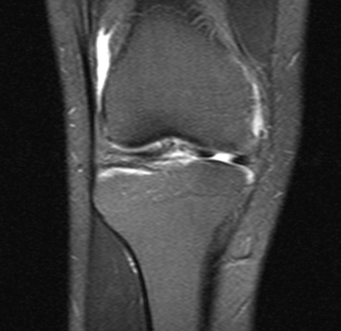
Two main arthroscopic techniques are used for meniscal tears: meniscal repair (suture fixation) and partial meniscectomy (limited removal of the damaged tissue). Both are performed arthroscopically through small incisions (approximately 1 cm); the choice depends on the location, type and vascular zone of the tear.
Arthroscopic Meniscal Repair — General Information
Arthroscopic meniscal repair is a technique in which the torn portion of the meniscus is first prepared to promote healing and then repaired with sutures, preserving the meniscal tissue. The knee is entered arthroscopically through small incisions (approximately 1 cm); the tear is visualised, its edges are debrided and it is repaired with sutures or implants.
- Aim: to preserve as much meniscal tissue as possible
- Duration: 30–60 minutes; general or spinal anaesthesia
- Discharge: usually the following day
- Complications: 1.3%; most are mild and temporary
Repair Techniques
- All-inside: entirely arthroscopic, using absorbable implants. The most commonly used technique.
- Inside-out: an additional small incision provides secure suture placement in large tears.
- Outside-in: used for tears of the anterior portion.
- Root repair: a tunnel is created in the tibia and the meniscal root is reattached.
Arthroscopic Partial Meniscectomy — General Information
The knee is entered arthroscopically through two small incisions (approximately 1 cm). While the damaged tissue is removed, healthy meniscal tissue is preserved to the maximum extent — a principle that is critical for long-term knee health.
- Aim: to preserve all healthy meniscal tissue possible
- Duration: 30 minutes; general or spinal anaesthesia
- Discharge: usually the following day
- Complications: 1.3%; most are mild and temporary
Discharge — Meniscal Repair
Patients are usually discharged after one or two nights in hospital. On the day after surgery you are mobilised with the physiotherapist and begin walking with a frame or crutches. You must not place any weight on the operated leg.
A knee brace is applied in theatre and must not be removed for 6 weeks. The angles of the brace are increased by your doctor when appropriate. On the first day the physiotherapist teaches closed-chain lower limb exercises, quadriceps and hamstring strengthening, and ankle pumping; it is important to perform these 3–4 times a day.
Discharge — Partial Meniscectomy
Patients are usually discharged after one night in hospital. On the day after surgery you are mobilised with the physiotherapist and begin walking with a frame. A frame is not required in every patient; it is generally used in older patients.
On the first day the physiotherapist teaches closed-chain lower limb exercises, quadriceps and hamstring strengthening, and ankle pumping; it is important to perform these 3–4 times a day.
Discharge Information Common to Both Procedures
Ice for 10–15 minutes four times a day is recommended after discharge. The medication prescribed includes an anticoagulant, which must be taken for 3 weeks. Compression stockings must be worn on the operated leg for 3 weeks and on the other leg for 1 week.
Swelling and pain are expected in the first 24–48 hours. Walking may begin the same day or the following day; after partial meniscectomy most patients do not require crutches. Strengthening and range-of-motion exercises begin from the first week within a pain-free range, and quadriceps and hamstring strengthening intensifies in weeks 1–3.
Early Postoperative Period — Factors Monitored
- Wound monitoring: discharge, marked redness and increased warmth are monitored for signs of infection
- Range of motion: being able to straighten the knee fully and a steady increase in flexion are very important. After a repair, the knee brace is worn for 6 weeks with the angle adjusted by your doctor.
- Pain: a steady reduction in pain is normal. Unless soft-tissue swelling develops around the knee, pain has usually settled within a week.
- Deep vein thrombosis: the formation of a clot in the deep veins of the leg, diagnosed by Doppler ultrasound. It is very rare (approximately 1 in 10,000) and is prevented by compression stockings and anticoagulation.
Early Period After Meniscal Repair (First 3 Weeks)
On the day after surgery you are mobilised with the physiotherapist wearing the knee brace and begin walking without placing weight on the operated leg. You are taught to walk with a frame or crutches. Your physiotherapist will then show you a set of exercises to be performed 4–5 times a day.
Weekly review is recommended, at which your exercises are checked, your dressings changed and your medication prescribed. It is essential to wear your compression stockings and take your anticoagulant during these first 3 weeks.
Early Period After Partial Meniscectomy (First 3 Weeks)
On the first postoperative day you are mobilised with the physiotherapist and shown a set of exercises. Unless microfracture or drilling was performed for cartilage damage, you may begin weight bearing and walking immediately. Crutches or a frame are usually not required (they are occasionally needed in older patients).
Weekly review is recommended, at which your exercises are checked, your dressings changed and your medication prescribed. It is essential to wear your compression stockings and take your anticoagulant during these first 3 weeks.
Risks & Complications
Both arthroscopic meniscal repair and partial meniscectomy are safe and effective procedures. The overall complication rate is around 1.3%, and most complications are mild and temporary.
Common
- Wound infection (recognised by discharge, marked redness and increased warmth)
- Swelling and pain in the first 24–48 hours after surgery (expected and temporary)
- Joint stiffness — resolves completely with physiotherapy (particularly after meniscal repair, because of the knee brace)
Risks Specific to Meniscal Repair
- Temporary bone loss: may develop because full weight bearing is not permitted for 6 weeks; it is entirely reversible and does not occur in every patient
- Early re-tear: bending the knee too far may cause the repair to fail in the early period, which is why the brace angles are increased gradually by the doctor
- Joint stiffness: may develop because knee flexion is limited to a set angle, but resolves completely with physiotherapy
Risks Specific to Partial Meniscectomy
- Risk of cartilage wear: depending on the amount of tissue removed there is a 13.5% likelihood at 5 years; this can be reduced by weight control and strengthening of the muscles around the knee
- Where microfracture or drilling is performed, weight bearing must be avoided for 4–6 weeks (this is done if cartilage damage is found during surgery)
Less Common
- Deep vein thrombosis (DVT): the formation of a clot in the deep veins of the leg; despite compression stockings and anticoagulation it may occur in approximately 1 in 10,000 cases. It is diagnosed by Doppler ultrasound and requires stronger anticoagulation and physiotherapy.
Long-term Risk
The risk of significant cartilage loss and osteoarthritis within 10–15 years is increased in patients who undergo total meniscectomy. This risk is lower after partial meniscectomy, and after meniscal repair it is reduced almost entirely. The surgeon therefore prefers to repair the meniscus with sutures whenever the tear is amenable to repair.
Contact Your Doctor Immediately If You Have
- Sudden increase in pain or swelling
- Discharge, marked redness or increased warmth at the wound
- Firm, one-sided swelling or redness of the leg (suspected DVT)
- A further episode of locking of the knee
- Unexpected loss of strength or impaired movement
Rehabilitation
The rehabilitation protocol differs markedly between the two procedures. After meniscal repair, weight bearing and knee flexion are advanced gradually to protect the sutures, whereas after partial meniscectomy early weight bearing and a rapid functional return are possible.
Rehabilitation After Meniscal Repair
- Days 1–3: Ice, elevation, compression. Knee brace set at 0–30°. Quadriceps and hamstring isometric exercises. No weight bearing on the operated leg.
- Days 3–21: Range of motion 0–30°. Still no weight bearing. The brace is continued and exercises are maintained.
- Weeks 4–6: Physiotherapy may begin and exercises continue. Light weight bearing may be permitted, depending on the type and location of the tear. The brace is continued.
- Weeks 6–12: Full weight bearing. The brace is discontinued. Cycling and swimming are recommended. Deep squatting is prohibited.
- Weeks 12–16: Proprioception and balance training. Straight-line running begins.
- Months 4–6: Sport-specific drills; pivoting and cutting movements.
Meniscal Repair — Criteria for Return to Sport
- Quadriceps strength at 90% of the opposite leg
- Full, pain-free range of motion
- 90% symmetry on the single-leg hop test
- No swelling or pain after activity
Rehabilitation After Partial Meniscectomy
- Phase 1 (days 1–3): Ice and elevation, pain control, weight bearing as tolerated, quadriceps and hamstring activation
- Phase 2 (days 3–21): Full weight bearing, gait normalisation, closed kinetic chain strengthening
- Phase 3 (weeks 3–6): Proprioception, balance training, swimming; return to desk-based work in this period
- Phase 4 (weeks 6–12): Running, lateral movements, sport-specific drills; return to sport once the criteria are met
Partial Meniscectomy — Criteria for Return to Sport
- Quadriceps and hamstring strength at least 90% of the opposite leg
- Full joint movement: pain-free flexion and extension
- Functional tests: single-leg hop, double jump test
- No swelling: no oedema after activity
- Average return to sport is 6 weeks; the decision should be based on criteria rather than the calendar and may vary between 6 and 8 weeks depending on sporting level
Knee Health After Repair
A successful meniscal repair preserves the meniscal tissue and supports long-term knee health. The following measures reduce the risk of osteoarthritis:
- Healthy weight: every additional kilogram places four times that load on the knee
- Quadriceps and hamstring strengthening
- Low-impact sports: swimming, cycling, elliptical trainer
- Avoid high-impact and pivoting sports (particularly for the first 12 months)
Lifestyle Advice
- Warm up before exercise and stretch afterwards
- Avoid squatting and kneeling for the first 4 months
- Descend stairs slowly and under control
- Attend your follow-up appointments
Is This Treatment Right for You?
Every patient's condition is different. After a detailed examination and imaging, the treatment plan that suits you best will be prepared.
Would you like more information about Meniscus Tears?
Book Appointment