Knee Cartilage Damage and Biological Treatments
Microfracture, mosaicplasty, PRP, hyaluronic acid and stem cell treatments for cartilage lesions
Overview
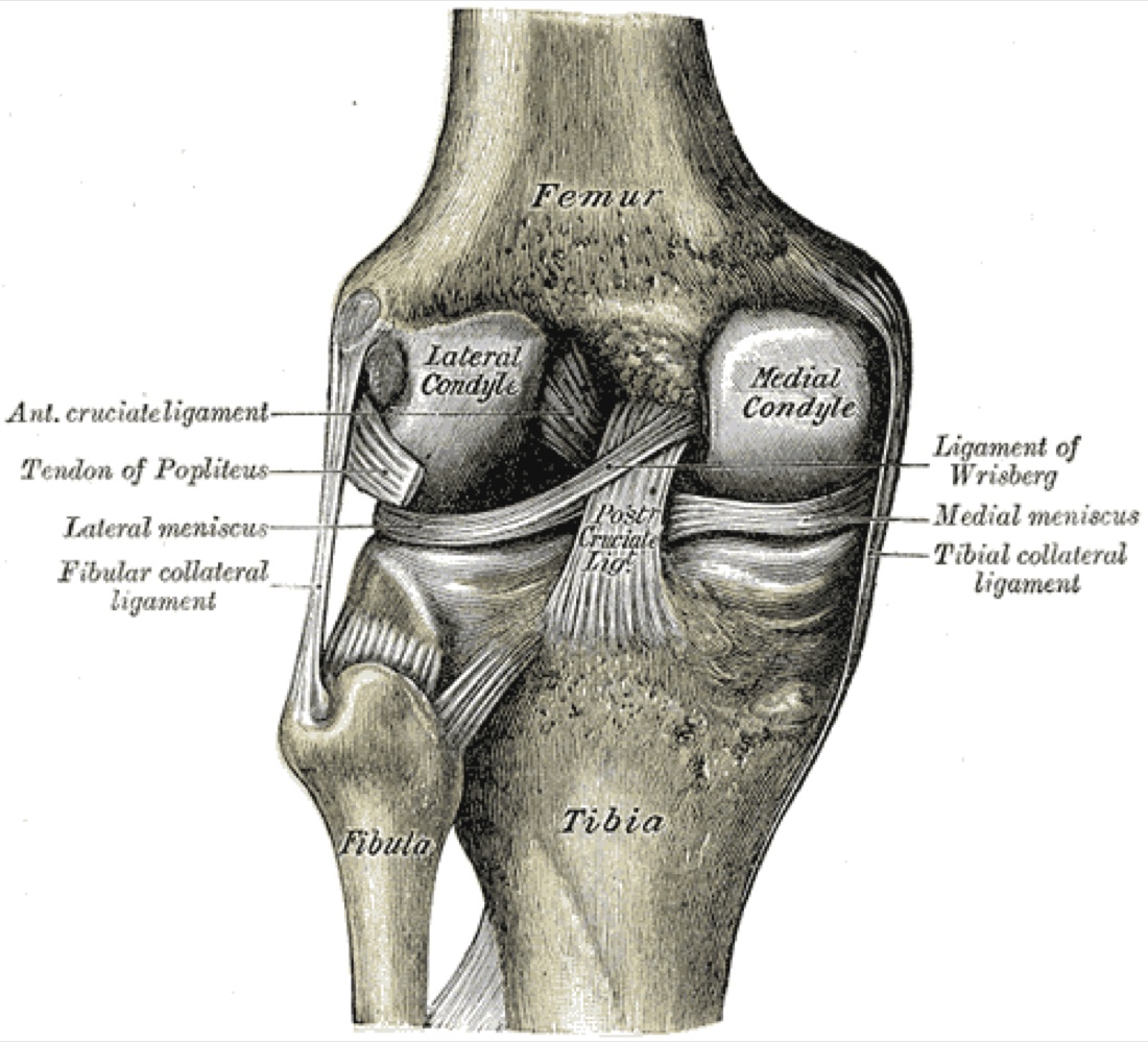
Cartilage damage in the knee is the breakdown of the smooth hyaline cartilage covering the joint surfaces, as a result of injury or degeneration. Because cartilage has no blood vessels of its own, its capacity to heal is very limited; untreated damage does not repair itself and may enlarge over time, progressing to knee osteoarthritis. Treatment options range widely according to the size and depth of the lesion, from intra-articular injections and biological treatments to cartilage repair surgery.
Articular Cartilage and the Healing Problem
The hyaline cartilage covering the joint surfaces is a specialised tissue that reduces friction almost to zero and spreads load across the whole surface. It contains no blood vessels, nerves or lymphatics; it is nourished entirely by diffusion from the joint (synovial) fluid.
This has two consequences: cartilage damage may cause no pain at first (there are no nerves), and once damaged, cartilage cannot repair itself (there is no blood supply). Every cartilage treatment is, in essence, an attempt to overcome this fundamental lack of healing capacity.
Cartilage damage affects not only the cartilage but also the subchondral bone beneath it. In advanced lesions, bone oedema, cyst formation and sclerosis develop.
Causes of Cartilage Damage
- Acute injury: Twisting, impact or a fall; often accompanies an anterior cruciate ligament or meniscal injury
- Repetitive microtrauma: In athletes and in occupations involving heavy loading
- Osteochondritis dissecans (OCD): Separation of a cartilage-bone fragment due to impaired blood supply to the subchondral bone, particularly in young patients
- Malalignment: Varus (bow-legged) or valgus (knock-kneed) alignment concentrates load on a narrow area of the joint
- Meniscal loss: Load on the cartilage rises markedly in knees where the meniscus has been removed
- Instability: Cartilage damage progresses over time in knees with anterior cruciate ligament deficiency
- Degeneration: The water content and elasticity of cartilage decrease with age
Outerbridge Classification
The depth of cartilage damage is decisive in choosing treatment. The most widely used arthroscopic grading is:
- Grade 0: Normal cartilage
- Grade 1: Softened, swollen cartilage with an intact surface
- Grade 2: Surface fissuring and fibrillation not reaching half the cartilage thickness
- Grade 3: Deep fissuring beyond half thickness, not reaching subchondral bone
- Grade 4: Full-thickness loss with exposed subchondral bone
Lesion Size and Treatment Choice
- Small lesion: <2 cm² — bone marrow stimulation (microfracture) is usually sufficient
- Medium lesion: 2 - 4 cm² — mosaicplasty (OAT), AMIC or cell-based treatments are considered
- Large lesion: >4 cm² — autologous chondrocyte implantation (ACI/MACI) or osteochondral allograft
- Location also matters: Lesions in the weight-bearing femoral condyle are managed differently from patellofemoral lesions
- Cartilage surgery performed without correcting associated malalignment or instability fails
Symptoms
- Deep knee pain that increases with activity: Particularly on stairs and when squatting
- Swelling: Joint effusion, especially after activity
- Catching or locking: When a loose cartilage fragment (loose body) is present
- Clicking and grinding (crepitus): As the knee bends and straightens
- Stiffness after prolonged sitting and pain with the first steps
- Feeling of instability: When there is an associated ligament injury
- No symptoms in the early stage: Because cartilage has no nerves, damage may progress silently
Diagnosis
The diagnosis is made from the history, examination and imaging. Injury history, duration of symptoms, activity level, previous knee surgery and associated conditions are reviewed. Examination assesses joint-line tenderness, effusion, range of movement, alignment, ligament stability and patellofemoral findings.
Plain radiographs (standing weight-bearing AP, lateral and patellofemoral views) do not show cartilage directly but provide information on joint-space narrowing, osteophytes and alignment. A long-leg radiograph is needed to measure the mechanical axis.
MRI is the main method for assessing cartilage, showing the size and depth of the lesion, subchondral bone oedema and associated meniscal and ligament pathology. Cartilage-specific sequences (T2 mapping, dGEMRIC) can detect early degeneration. Arthroscopy offers both definitive diagnosis and treatment; the lesion is seen directly and graded.
Non-operative and Biological Treatments
Treatment is stepwise. In early-stage and small lesions, and in patients unsuitable for surgery, non-operative methods are tried first. Biological treatments are an important part of this step; their aim is not to restore damaged cartilage, but to reduce pain, improve the joint environment and slow progression.
- Activity modification and weight control: Every extra kilogram is multiplied as load on the knee; weight loss is the most effective non-operative measure
- Physiotherapy: Strengthening of the quadriceps and hip muscles, range of movement and proprioception
- Analgesics and anti-inflammatory medication (NSAIDs): Short-term, under medical supervision
- Hyaluronic acid injection (viscosupplementation): Supports the lubricating and shock-absorbing properties of joint fluid; used particularly for pain control in early to moderate degeneration. The effect is not permanent and can be repeated at intervals
- PRP (platelet-rich plasma): Plasma rich in platelets and growth factors, separated from the patient's own blood by centrifugation, is injected into the joint. Because the patient's own tissue is used, there is no risk of allergy or rejection
- Mesenchymal stem cell treatments: Cells obtained from fat tissue or bone marrow are injected into the joint. This field is still under research; studies on effectiveness and long-term results are ongoing
- Corticosteroid injection: Gives short-term relief when there is marked inflammation and effusion; used sparingly because frequent repetition may harm cartilage
- Knee brace and insoles: In malaligned knees, may shift load to the healthy compartment
- Realistic expectations of biological treatments: These methods do not regenerate damaged cartilage. They may reduce pain and improve function; the response varies from patient to patient
Surgical Options
Surgery is considered in patients who do not respond to non-operative treatment, who have mechanical symptoms (locking, catching) and who have a significant lesion. The method is chosen according to lesion size and depth and the patient's age and activity level.
- Arthroscopic debridement and loose body removal: Clearing loose fragments; gives relief in patients with mechanical symptoms
- Microfracture / nanofracture (bone marrow stimulation): In small lesions, stem cells from holes made in the subchondral bone form fibrocartilage
- AMIC (autologous matrix-induced chondrogenesis): A collagen membrane placed over microfracture holds the cells in place
- Mosaicplasty / OAT: Cylinders of cartilage and bone taken from a non-weight-bearing area of the knee are transferred into the lesion; a true hyaline cartilage transfer
- ACI / MACI (autologous chondrocyte implantation): The patient's own cartilage cells are grown in the laboratory and implanted into the lesion; a two-stage treatment
- Osteochondral allograft: Reconstruction of very large, deep lesions with a donor cartilage-bone block
- Correction of associated problems: Osteotomy for malalignment, ligament reconstruction for instability, meniscal repair for meniscal loss — if neglected, cartilage surgery fails
Expected Outcomes
The outcome of cartilage treatment depends on lesion size and location, age, weight and whether associated problems have been corrected. After microfracture of small lesions there is marked relief in the short to medium term; however, because the fibrocartilage formed is weaker than hyaline cartilage, symptoms may return in the long term.
Long-term results are better with mosaicplasty and cell-based treatments, but recovery is longer. Biological injections (PRP, hyaluronic acid) may help with pain control; they should not be expected to regenerate cartilage. No method replaces joint replacement in advanced, widespread osteoarthritis.
Surgery & First 3 Weeks
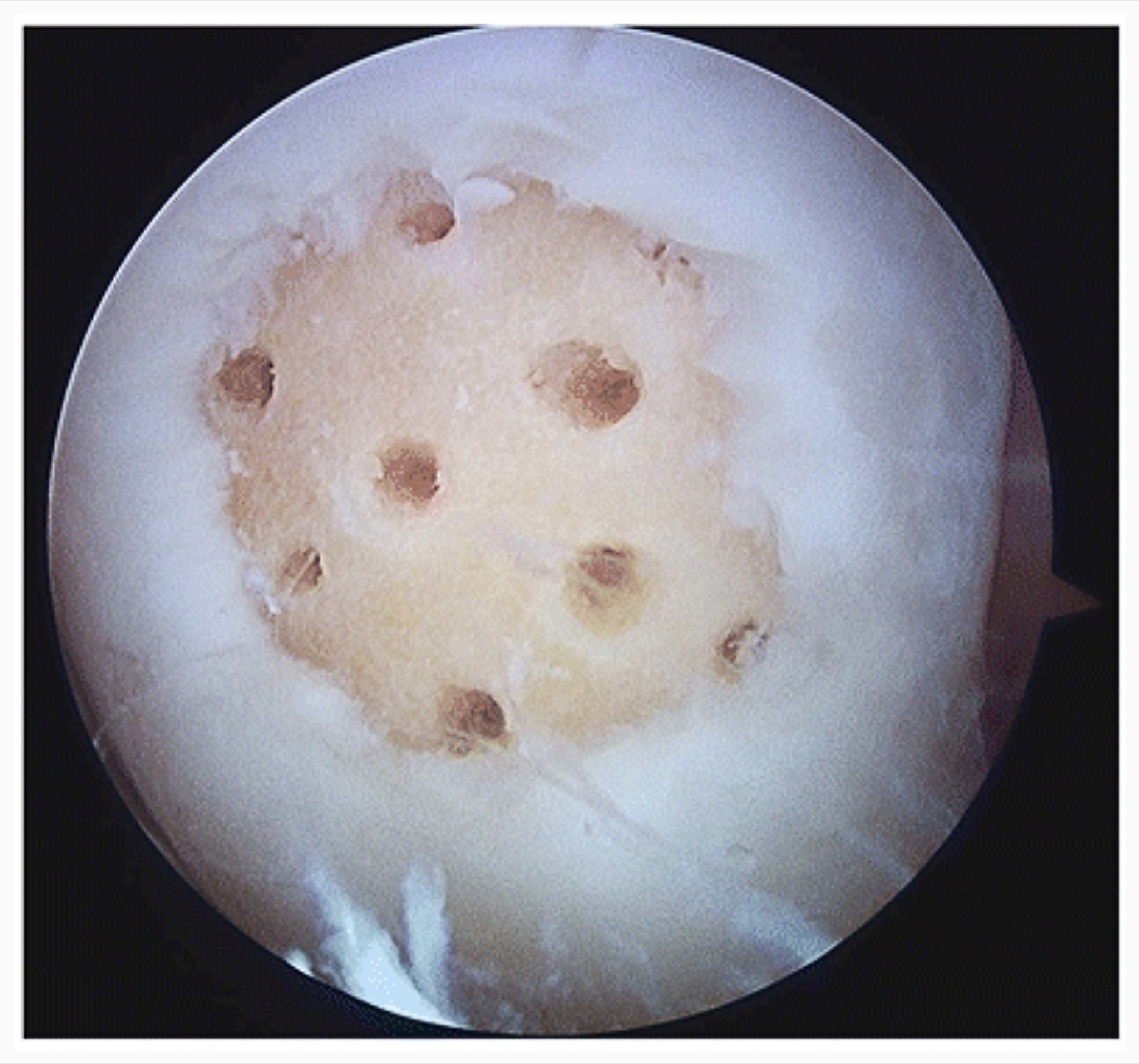
Knee cartilage surgery is performed under spinal/epidural or general anaesthesia. Arthroscopic procedures can usually be done as day cases, while more extensive procedures such as mosaicplasty and cell-based treatments may require 1 - 2 days in hospital.
General Information About the Operation
Radiographs, a long-leg film and MRI are reviewed for planning. If malalignment or ligament deficiency is found, correction is planned in the same session or beforehand. Prophylactic antibiotics are given.
- Duration: Arthroscopic debridement / microfracture 30 - 60 minutes; mosaicplasty 90 - 150 minutes; ACI/MACI second stage 90 - 120 minutes
- Anaesthesia: Spinal/epidural or general
- Hospital stay: Arthroscopic cases day case to one night; open and combined cases 1 - 2 days
Intra-articular Injections
- Performed in clinic under sterile conditions; no operating theatre is needed
- PRP: 15 - 60 ml of blood is taken, platelet-rich plasma is separated by centrifugation and injected into the joint. Usually 1 - 3 sessions, weeks apart
- Hyaluronic acid: May be given as a single dose or as 3 - 5 weekly injections, depending on the preparation
- Stem cell treatments: Cells obtained from fat tissue (by liposuction) or bone marrow (aspirated from the iliac crest) are processed and injected
- Ultrasound guidance ensures the needle reaches the right place
- Strenuous activity is avoided and ice applied for 24 - 48 hours after injection
- A temporary increase in pain in the first days after PRP is expected
Arthroscopic Debridement and Loose Body Removal
- Camera and instruments are introduced through anteromedial and anterolateral portals
- Loose cartilage or bone fragments are removed
- Unstable, hanging cartilage flaps are trimmed and the lesion edges smoothed
- Any associated meniscal tear is treated in the same session
- Gives marked relief in patients with mechanical symptoms (locking, catching); not sufficient alone in widespread osteoarthritis
Microfracture / Nanofracture (Bone Marrow Stimulation)
The most widely used cartilage repair technique. Indication: full-thickness (Grade 3 - 4) lesions smaller than 2 cm².
- The lesion edges are debrided back to healthy cartilage, creating vertical walls (critical for the new tissue to stay in place)
- The calcified cartilage layer is gently removed with a curette
- Holes 2 - 4 mm deep are made in the subchondral bone at 3 - 4 mm intervals using a special awl or K-wire
- Blood and marrow cells from the holes form a clot on the lesion base, which gradually turns into fibrocartilage
- Advantage: A single-stage, short procedure requiring no additional materials
- Limitation: The fibrocartilage formed is biomechanically weaker than hyaline cartilage
AMIC — Matrix-assisted Microfracture
- After microfracture, a collagen or hyaluronic membrane is placed over the lesion
- The membrane holds the marrow-derived cells in place, supporting the formation of better-quality tissue
- It is single-stage and requires no cell culture, making it more practical than ACI
- Preferred for medium-sized (2 - 4 cm²) lesions or where microfracture alone would be insufficient
Mosaicplasty / OAT (Osteochondral Autograft Transfer)
The technique in which true hyaline cartilage is transferred together with its subchondral bone. Indication: lesions of 2 - 4 cm², particularly where subchondral support is compromised.
- Donor site: Cylindrical cartilage-bone grafts 6 - 10 mm in diameter are taken from non-weight-bearing areas of the knee (trochlear ridge or near the intercondylar notch)
- Recipient site: Cylindrical tunnels of the same diameter are drilled into the lesion base
- The grafts are press-fitted so that their surfaces sit exactly level with the surrounding cartilage — a graft that sits proud or recessed compromises the result
- Several cylinders are placed side by side to cover the lesion like a mosaic
- Can be performed arthroscopically or through a mini-open approach, depending on lesion location
ACI / MACI — Autologous Chondrocyte Implantation
- Stage 1: A small cartilage biopsy is taken arthroscopically from a non-weight-bearing area; the cells are grown in the laboratory for 4 - 6 weeks
- Stage 2: The lesion is prepared through an open or mini-open procedure; the cultured chondrocytes are placed beneath a periosteal flap (classic ACI) or on a collagen membrane (MACI)
- Advantage: Formation of hyaline-like cartilage; long-term results in large lesions are promising
- Disadvantage: Two-stage surgery, high cost and long rehabilitation
Correction of Associated Problems
The success of cartilage surgery depends on correcting the underlying cause of the lesion. If this step is skipped, the cartilage repair soon breaks down.
- Malalignment: High tibial osteotomy for varus, distal femoral osteotomy for valgus, shifting load to the healthy compartment
- Anterior cruciate ligament deficiency: ACL reconstruction in the same session or beforehand
- Meniscal loss: Meniscal repair where possible; meniscal transplantation in selected cases
- Patellofemoral instability: MPFL reconstruction or tibial tubercle transfer
Discharge and Weight-bearing Protocol
Arthroscopic procedures are usually day cases. The postoperative weight-bearing protocol differs markedly depending on the method used, and adherence directly affects the result. Newly formed cartilage tissue collapses under early load.
- Arthroscopic debridement: Weight-bearing as tolerated
- Microfracture / AMIC: 6 - 8 weeks partial or non-weight-bearing (depending on lesion location)
- Mosaicplasty / OAT: 6 - 8 weeks non-weight-bearing
- ACI / MACI: 6 - 8 weeks non-weight-bearing, then gradual progression
- Patellofemoral lesions: Weight-bearing may start earlier but knee flexion is limited with a brace
- Continuous passive motion (CPM): Recommended for several hours a day to support cartilage nutrition and healing
- Ice and elevation 3 - 4 times a day for 15 - 20 minutes. Analgesics, anti-inflammatory medication, stomach protection and anticoagulation until weight-bearing are prescribed. Stitches are removed at 10 - 14 days
Early Postoperative Period (First 3 Weeks)
The aim in this period is to keep the joint moving while protecting the new cartilage tissue from load. Cartilage is nourished by movement but harmed by load; the whole logic of rehabilitation rests on this dilemma.
- Strict adherence to the weight-bearing protocol: Early loading causes the repair tissue to collapse and the surgery to fail
- Early passive movement: CPM or manual passive knee movement directly supports cartilage nutrition and healing quality
- Quadriceps isometric exercises: 3 - 4 times a day to slow muscle wasting
- Ankle pumps and hip exercises: Reduce swelling and the risk of DVT
- Swelling control: Ice, elevation and compression stockings
- Wound care: Portal sites kept dry for 10 - 14 days; discharge, redness and warmth monitored
- Thromboprophylaxis: Anticoagulation and compression stockings; the risk is higher during non-weight-bearing
- After injections: Avoid strenuous activity for 24 - 48 hours; a temporary increase in pain is expected
Follow-up
- Days 10 - 14: Removal of stitches
- Week 6: Weight-bearing decision and progression of rehabilitation
- Month 3: Strength and range-of-movement assessment
- Month 6 and year 1: Follow-up MRI if needed
Risks & Complications
Cartilage treatments, particularly arthroscopic procedures and injections, are safe and serious complications are rare. However, this field has one particular reality: no method can restore damaged hyaline cartilage exactly, and results vary considerably between patients.
Risks Specific to Injection Treatments
- Temporary increase in pain and swelling: Particularly in the first 2 - 3 days after PRP; this is expected
- Joint infection (septic arthritis): Very rare but the most serious risk; strict sterile technique is essential
- Temporary tenderness and bruising at the injection site
- With corticosteroid injection: Raised blood glucose (in diabetes), thinning of subcutaneous fat, skin lightening; frequent repetition may harm cartilage
- Flare reaction after hyaluronic acid (pseudoseptic reaction): Rare
- Effectiveness: These treatments do not regenerate cartilage; failure to respond is not a complication but a known possibility
- Stem cell treatments: Long-term effectiveness and safety data are still accumulating; this uncertainty should be explained clearly to the patient
Surgical Risks — Common
- Superficial portal infection (usually treated with antibiotics)
- Postoperative pain, swelling and joint effusion
- Temporary numbness or tenderness at the portal sites
- Temporary knee stiffness and restricted movement — improves with physiotherapy
- Quadriceps wasting: Develops markedly during non-weight-bearing
- Prolonged swelling: May last 6 - 12 months
Surgical Risks — Less Common
- Deep joint infection (septic arthritis): Under 1%; requires urgent arthroscopic washout and antibiotics
- Deep vein thrombosis (DVT) and pulmonary embolism: Increased risk during non-weight-bearing
- Haemarthrosis (bleeding into the joint)
- Nerve injury: Injury to the infrapatellar branch of the saphenous nerve may cause numbness on the front-inner knee
- Arthrofibrosis (intra-articular adhesions): Marked restriction of movement; occasionally requires arthroscopic release
- Complex regional pain syndrome (CRPS)
Method-specific Risks
- Microfracture: Wear of the fibrocartilage over time with return of symptoms; intralesional osteophyte formation; subchondral cystic degeneration
- Mosaicplasty / OAT: Pain and swelling at the donor site (donor-site morbidity); graft subsidence; mismatch between graft surface and surrounding cartilage; gaps between cylinders
- ACI / MACI: Periosteal or membrane hypertrophy (may need debridement); graft delamination; need for two-stage surgery
- Allograft: Delayed or failed graft integration; limited tissue availability; immune reaction (rare)
Risk Factors
- Obesity: Directly affects both the surgical result and the load on the cartilage
- Advanced age: Cartilage healing capacity falls markedly after 40 - 50
- Smoking: Impairs tissue healing
- Poorly controlled diabetes
- Uncorrected malalignment or ligament deficiency: The most common cause of failed cartilage surgery
- Widespread (bipolar) damage: Damage to both opposing surfaces markedly worsens the outcome
- Inflammatory joint disease (rheumatoid arthritis, etc.)
- Poor adherence to the weight-bearing protocol
Contact Your Doctor Immediately If
- Pain, swelling or warmth in the knee suddenly increases
- You have a temperature above 38 °C
- There is discharge, a bad smell or an opening in the wound
- There is firm, one-sided swelling and redness of the leg (suspected DVT)
- You develop shortness of breath or chest pain (suspected pulmonary embolism)
- The knee suddenly locks or cannot be moved
- Numbness, tingling or weakness in the leg is progressing
Long-term Outcomes
Most patients with small lesions report marked relief in the short and medium term after microfracture; however, symptoms may return in the long term as the fibrocartilage wears. Long-term durability is better with mosaicplasty and cell-based treatments.
Progression of cartilage damage to osteoarthritis cannot always be prevented, even with treatment; the risk is highest with large, bipolar lesions and uncorrected malalignment. Knee replacement is considered when advanced, widespread osteoarthritis develops. Weight control, maintaining muscle strength, correcting associated problems and avoiding high-impact activity are the key determinants of long-term success.
Rehabilitation
Rehabilitation after cartilage surgery is one of the longest and most demanding programmes in orthopaedics. The core principle is: early movement, late load. Movement nourishes the cartilage and improves healing quality; early load collapses the newly formed tissue. The programme differs markedly according to the method used and the location of the lesion.
After Microfracture / AMIC
- Weeks 0 - 2: Non-weight-bearing or partial weight-bearing (crutches). CPM or manual passive movement for several hours a day. Quadriceps isometrics, ankle pumps. Ice and elevation.
- Weeks 2 - 6: Non-weight-bearing continues. Passive and active-assisted range of movement increased. Quadriceps and hamstring isometrics. Stationary cycling (no resistance, for movement).
- Weeks 6 - 8: Gradual weight-bearing (partial → full). Weaning off crutches. Active range of movement and light strengthening.
- Months 2 - 4: Full weight-bearing; gait normalises. Closed-chain exercises (mini squats, limited-arc leg press). Balance and proprioception. Swimming.
- Months 4 - 6: Strengthening intensifies. Light running on flat ground (with the surgeon's approval).
- Months 6 - 12: Gradual return to sport-specific drills; high-impact activity at 9 - 12 months. Fibrocartilage takes about 12 months to mature.
After Mosaicplasty / OAT
- Weeks 0 - 6/8: Non-weight-bearing. Early passive movement is critical. A separate exercise programme is followed for the donor site.
- Weeks 8 - 12: Gradual weight-bearing. Active movement and light strengthening. Stationary cycling, pool walking.
- Months 3 - 6: Full weight-bearing; strengthening and proprioception intensify. Light running begins (around months 5 - 6).
- Months 6 - 9: Sport-specific work.
- Months 9 - 12: Full return to high-impact activity with the surgeon's approval.
After ACI / MACI
- Weeks 0 - 6: Non-weight-bearing; CPM recommended. Early movement is critical for maturation of the implanted cells.
- Weeks 6 - 12: Gradual weight-bearing. Active range of movement and light strengthening.
- Months 3 - 6: Full weight-bearing; return to low-impact activity. Strengthening and balance.
- Months 6 - 12: Gradual return to sport.
- Months 12 - 18: Full maturation of the cartilage; return to high-impact activity.
Special Notes for Patellofemoral Lesions
- Weight-bearing can often begin earlier, but knee flexion is limited with a brace
- For the first 6 weeks, walking is usually allowed with controlled flexion of 0 - 30° / 0 - 60°
- Deep squatting, going downstairs and prolonged sitting with the knee bent are limited for the first 3 months
- Quadriceps work is done at low angles and low resistance
Rehabilitation After Injection Treatments
- First 24 - 48 hours: Avoid strenuous activity and apply ice. A temporary increase in pain is expected
- From week 1: Return to normal daily activities
- Continue a regular quadriceps and hip strengthening programme — this is the most important factor in prolonging the effect of the injection
- Low-impact activities (swimming, cycling, walking) are encouraged
- Weight control: Directly affects the effectiveness of injection treatment
Return-to-sport and Activity Criteria
- Quadriceps strength at least 85 - 90% of the other leg
- Full, pain-free knee range of movement
- No swelling or pain after activity
- 90% symmetry on single-leg hop tests
- Pain-free completion of sport-specific movements
- Sufficient time elapsed for the method used (cartilage maturation takes time)
- Approval from the surgeon and physiotherapist; follow-up MRI if needed
Recommended and Avoided Activities
- Recommended: Swimming, stationary and outdoor cycling, elliptical trainer, walking on flat ground, water exercise
- Recommended: Low-impact strengthening, Pilates, controlled yoga
- Avoid (especially in the first year): Jumping and high-impact activities
- Avoid: Deep squatting and heavy squats
- Avoid: Sports requiring pivoting and sudden changes of direction (football, basketball, tennis)
- Avoid: Running on uneven ground and long-distance running
Lifestyle and Recovery Advice
- Follow the weight-bearing programme to the letter — in this surgery, early loading is the most common cause of failure
- Do your home exercise programme every day; passive movement is critical for cartilage nutrition
- Weight control: Every 1 kg of excess weight becomes 3 - 4 kg of extra load on the knee when walking. Losing weight is the strongest factor in prolonging the effect of cartilage treatment
- Do not smoke; it markedly impairs tissue healing
- Maintain quadriceps strength for life; strong muscles reduce the load on cartilage
- If you have diabetes, keep your blood glucose tightly controlled
- Do not postpone correction of malalignment or ligament deficiency; without it, cartilage treatment is short-lived
- Be patient: Cartilage healing is measured in months, approaching a year
- Attend regular follow-up (week 6, month 3, month 6, year 1)
What to Expect
After microfracture, return to daily activities takes 3 - 4 months and return to sport 6 - 12 months. These times are longer after mosaicplasty (6 - 9 months for daily activities, 9 - 12 months for sport). Full recovery after ACI/MACI may take 12 - 18 months.
Mild swelling, stiffness and discomfort after activity may persist for the first 6 - 12 months; this is expected and settles with time. After injection treatments, the effect usually begins within a few weeks and its duration varies; it is not permanent. The key determinants of outcome are adherence to the weight-bearing programme, weight control and correction of associated problems.
Is This Treatment Right for You?
Every patient's condition is different. After a detailed examination and imaging, the treatment plan that suits you best will be prepared.
Would you like more information about Knee Cartilage Damage and Biological Treatments?
Book Appointment