Distal Biceps Tendon Rupture
Surgical repair of rupture of the biceps tendon at its insertion on the elbow
Overview
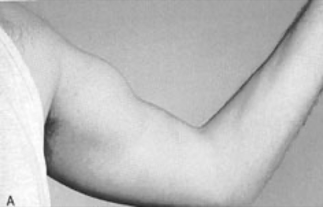
Distal biceps tendon rupture is the detachment of the biceps muscle from its insertion at the elbow (the radial tuberosity). It typically occurs in men aged 40 - 60, in the dominant arm, when an unexpected, sudden load is applied to a bent elbow. The biceps not only bends the elbow but is also the main source of forearm supination (turning the palm up). In untreated ruptures, the most noticeable loss is therefore strength in movements such as turning a door handle or using a screwdriver. Surgical repair gives reliable results, particularly in active patients and when performed early.
Anatomy of the Distal Biceps
The biceps brachii lies at the front of the upper arm and arises from the shoulder by two heads (long and short). At the elbow it forms a single tendon that inserts on the radial tuberosity, a bony prominence at the upper end of the radius.
The muscle has two main functions: bending the elbow (flexion) and turning the forearm outwards (supination). About 40 - 50% of supination strength and 25 - 30% of flexion strength comes from the biceps. When the tendon ruptures and retracts, these strengths fall considerably, but other muscles (brachialis, brachioradialis, supinator) take over, so the patient can still perform the movement — which is why the rupture can be missed.
The lacertus fibrosus, a sheet-like band from the tendon, may prevent retraction in partial ruptures. An intact lacertus can hold a ruptured tendon near its position and create a misleading picture on examination.
The blood supply to the insertion zone is relatively poor, which forms the anatomical basis for rupture.
Mechanism and Risk Factors
- Typical mechanism: A sudden, unexpected eccentric load with the elbow bent at about 90° — a heavy object slipping while being lifted, or trying to catch a falling load
- Weight training: Particularly sudden strain during biceps curls
- Sex and age: Most ruptures occur in men aged 40 - 60, in the dominant arm
- Smoking: Impairs tendon blood supply and markedly increases the risk
- Anabolic steroid and corticosteroid use
- Fluoroquinolone antibiotics
- Pre-existing tendinopathy: Most ruptures occur in a tendon with a degenerative background
- Narrow space around the radial tuberosity: Impingement of the tendon in this space during forearm rotation contributes to degeneration
Symptoms
- Sudden sharp pain: At the front of the elbow; most patients describe hearing a 'pop' or feeling something tear
- Reverse Popeye deformity: The biceps bunches up towards the shoulder, creating a bulge in the upper arm, while the front of the elbow looks hollow
- Gap in the elbow crease: The normally palpable, taut tendon is no longer felt
- Bruising: At the front of the elbow and the inner forearm, becoming more obvious over the first days
- Loss of strength: The most noticeable loss is in supination — difficulty turning a door handle, using a screwdriver or opening a jar
- Fatigue-related pain: Tiring quickly with repeated lifting
- Cramping sensation at the front of the arm
- Misleading sign: The patient can still bend the elbow (the brachialis takes over), so the rupture may be underestimated
Diagnosis
The diagnosis is largely clinical. How the injury happened, whether a pop was heard, smoking and medication are reviewed. Early diagnosis matters because it directly determines the treatment options.
Examination looks for absence of the palpable tendon in the elbow crease, retraction of the biceps and bruising. The hook test is the most valuable sign: with the elbow bent to 90° and the forearm supinated, the examiner tries to hook an index finger under the biceps tendon from the lateral side; with an intact tendon the finger hooks under it, with a complete rupture it cannot. Supination strength is compared with the other arm; the biceps squeeze test is also used.
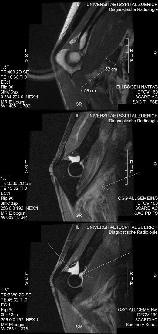
Ultrasound is quick and widely available and, in experienced hands, shows the rupture and the degree of retraction. MRI confirms the diagnosis, distinguishes partial from complete rupture, shows how far the tendon has retracted and the tissue quality, and is valuable for planning. A dedicated MRI with the forearm supinated and elbow flexed (FABS position) shows the insertion more clearly.
Plain radiographs do not show the rupture; they are taken to exclude a bony avulsion from the radial tuberosity or other bone pathology.
Non-operative Treatment
Non-operative treatment can be used in selected patients. Choosing it means accepting a permanent loss of strength, so the decision should be made after an open discussion with the patient.
- Typical candidates: Older, low-demand patients; those with a rupture in the non-dominant arm; those at high surgical risk; those who prefer not to have surgery
- Short-term splint or sling for comfort, followed by early movement
- Pain control with ice, elevation and anti-inflammatory medication
- A graded range-of-movement and strengthening programme
- Expected outcome: A permanent loss of about 30% of elbow flexion strength and about 40 - 50% of supination strength, with early fatigue in repetitive tasks
- The cosmetic deformity (Popeye appearance) is permanent
- Pain usually settles within weeks; many patients can continue their daily lives
Surgical Treatment
Surgical repair is the preferred treatment for active patients and those in manual or strength-demanding occupations. Timing is critical: in repairs performed within the first 3 - 4 weeks, the tendon has not yet retracted far and tissue quality is preserved, so anatomical fixation is possible.
- Acute repair (first 3 - 4 weeks): The tendon is reattached directly to the radial tuberosity; the best results are seen in this group
- Late / chronic cases (after 4 - 6 weeks): The tendon has retracted and shortened, so direct reattachment may not be possible; reconstruction with a graft may be needed
- Fixation methods: Cortical button, interference screw, suture anchors or bone tunnel techniques; often combined
- Surgical approach: Single-incision (anterior) or double-incision techniques
- The main advantage of surgery is recovery of most supination and flexion strength
Which Treatment for Which Patient?
- Young or middle-aged, active patient with a rupture in the dominant arm → surgical repair
- Heavy manual worker, technician, athlete or other strength-demanding occupation → surgical repair
- Older, low-demand patient with a rupture in the non-dominant arm → non-operative treatment is appropriate
- Serious comorbidities, high surgical risk → non-operative treatment
- Partial ruptures: Non-operative if symptoms are mild; surgery if loss of strength and pain persist
- The most important element of the decision is how much the patient needs supination strength in daily life
Expected Outcomes
After early surgical repair, most patients regain elbow flexion and supination strength close to the other arm. Return to daily activities takes about 3 months; return to heavy work and sport takes 4 - 6 months.
The cosmetic appearance is largely restored by surgical repair. Good results can also be achieved with graft reconstruction in chronic cases, but strength recovery may not be as complete as after acute repair and the complication rate is higher. In patients treated non-operatively, pain settles but the loss of strength is permanent.
Surgery & First 3 Weeks
Distal biceps tendon repair is performed under a regional block (axillary or supraclavicular), general anaesthesia or a combination of both. A regional block is effective for pain control in the first 12 - 24 hours. The patient lies supine with the arm on a side table and a tourniquet is used.
General Information About the Operation
Preoperative MRI is used to assess the degree of retraction and tissue quality; in chronic cases a graft is prepared. Prophylactic antibiotics are given.
- Duration: Acute repair 60 - 90 minutes; chronic / graft reconstruction 90 - 150 minutes
- Anaesthesia: Regional block, general anaesthesia or a combination
- Hospital stay: Usually day case or one night
- Timing: Surgery within the first 3 - 4 weeks is the aim where possible
Surgical Approaches
- Single-incision (anterior) approach: The radial tuberosity is reached through a 4 - 6 cm transverse or longitudinal incision in the elbow crease. Advantages: one incision, lower risk of heterotopic ossification. Disadvantage: higher risk of injury to the posterior interosseous nerve (PIN), a branch of the radial nerve
- Double-incision approach: The tendon is found through a small anterior incision, and the tuberosity is reached through a second posterolateral incision. Advantages: lower PIN risk, easier anatomical fixation. Disadvantage: higher risk of heterotopic ossification and radioulnar synostosis (a bony bridge joining the two forearm bones)
- The choice depends on the surgeon's experience and the features of the case; good results are reported with both techniques
Acute Repair — Surgical Technique
- An incision is made in the elbow crease; the lateral antebrachial cutaneous nerve under the skin is identified and protected (it is the structure most often injured)
- The ruptured tendon end is found — usually retracted up within the muscle sheath — and brought down
- The tendon end is debrided and prepared with strong locking sutures (Krackow or similar technique)
- The forearm is supinated to expose the radial tuberosity; this manoeuvre moves the posterior interosseous nerve away from the working area and is the most important safety step of the operation
- Soft tissue over the tuberosity is cleared and the bone surface prepared for tendon healing
- Fixation: A cortical button passed through the far cortex of the radius, supplemented if needed by an interference screw. Suture anchors or bone tunnels are alternatives
- Elbow range of movement and tendon tension are checked on the table after fixation
- Closure in layers; a drain is usually not needed
Reconstruction for Chronic and Late Cases
- If the tendon has remained retracted for more than 4 - 6 weeks, it shortens and cannot reach the tuberosity directly
- Graft options: Semitendinosus autograft, Achilles tendon allograft or flexor carpi radialis graft
- The graft bridges between the biceps tendon stump and the tuberosity
- The risk of nerve injury and heterotopic ossification is higher in these cases, and recovery is longer
- Strength recovery may not be as complete as after acute repair
Discharge Information
Patients usually go home the same day or the next day. After surgery the arm is protected in a splint or hinged elbow brace with the elbow at about 90° and the forearm in neutral to supination. This position reduces tension on the repair.
In the modern approach, if fixation is strong enough, early controlled movement is preferred; prolonged rigid immobilisation leads to elbow stiffness. The brace angle is opened gradually according to the surgeon's protocol.
Ice for 15 - 20 minutes three to four times a day and elevation of the arm above heart level are recommended. Analgesics, anti-inflammatory medication and stomach protection are prescribed. Some centres use anti-inflammatory medication for 2 - 4 weeks to prevent heterotopic ossification. Stitches are removed at 10 - 14 days.
Early Postoperative Period (First 3 Weeks)
The aims in this period are to protect the repair, control swelling, prevent elbow stiffness and monitor nerve function closely. Two risks pull in opposite directions in this operation: early strain disrupts the repair, while excessive immobility stiffens the elbow.
- NO active elbow flexion: Active biceps contraction (especially resisted flexion and supination) is strictly prohibited for the first 3 - 6 weeks — it pulls directly on the repair
- Passive and active-assisted movement: Gentle elbow bending and straightening with help from the good arm, according to the surgeon's protocol
- Extension limit: Full straightening is reached gradually over the first weeks; sudden, forced straightening stresses the repair
- Hand, wrist and shoulder exercises: 3 - 4 times a day; reduce swelling and prevent stiffness in neighbouring joints
- No lifting: Nothing is lifted with the operated arm for the first 6 weeks (even a glass of water is limited)
- Swelling control: Ice and elevation of the arm above heart level
- Wound care: Kept dry for 10 - 14 days; discharge, redness and warmth monitored
- Nerve monitoring: Numbness on the outer forearm (lateral antebrachial cutaneous nerve), weakness of thumb and index finger movements, or inability to lift the wrist and fingers (posterior interosseous nerve) must be reported immediately
Follow-up
- Days 10 - 14: Removal of stitches and review of brace settings
- Week 6: Decision on progressing to active movement and light strengthening
- Month 3: Strength assessment and decision on heavy activity
- Month 6: Assessment for return to sport and full strength
Risks & Complications
Distal biceps tendon repair is generally a successful operation with high patient satisfaction. However, because of the local anatomy it has specific risks: important nerves lie close to the radial tuberosity, and the area is prone to heterotopic ossification.
Risks Specific to Non-operative Treatment
- Permanent loss of strength: About 40 - 50% in supination and about 30% in elbow flexion
- Early fatigue and loss of endurance with repetitive movements
- Permanent cosmetic deformity (Popeye appearance)
- Cramp-like pain at the front of the arm
- If surgery is considered later, the tendon will have retracted and shortened, making a graft necessary and results more limited
Surgical Risks — Common
- Lateral antebrachial cutaneous nerve injury: The most common complication of this operation. Causes numbness or tingling on the outer forearm; usually temporary, permanent in some patients
- Postoperative pain, swelling and bruising
- Superficial wound infection (usually treated with antibiotics)
- Temporary elbow stiffness, particularly loss of full extension — improves with physiotherapy
- Scar sensitivity
Surgical Risks — Less Common
- Posterior interosseous nerve (PIN) injury: The motor branch of the radial nerve; injury causes inability to lift the fingers and wrist (wrist drop). The risk is higher with the single-incision technique. Usually temporary but may be permanent
- Heterotopic ossification: Bone formation in soft tissue; more common with the double-incision technique. May restrict movement
- Radioulnar synostosis: A bony bridge between the two forearm bones; severely restricts forearm rotation (supination and pronation). Rare but difficult to treat
- Repair failure (re-rupture): Usually due to not following the protocol or lifting weights early
- Fracture of the radial head or neck: Related to the drill tunnel (very rare)
- Deep infection (under 1%)
- Hardware irritation or displacement of the button
- Complex regional pain syndrome (CRPS): Rare
- Vascular (brachial artery) injury: Very rare
Risk Factors
- Delayed surgery (after 4 - 6 weeks): Both technical difficulty and complication rate increase
- Smoking: Markedly impairs tendon and wound healing
- Poorly controlled diabetes
- Anabolic steroid or corticosteroid use
- Obesity and a bulky arm (makes surgical exposure more difficult)
- Need for a graft in chronic cases
- Poor adherence to the postoperative protocol, particularly early lifting
- History of heterotopic ossification
Contact Your Doctor Immediately If
- Pain suddenly increases or the arm suddenly feels 'loose' (suspected repair failure)
- You have a temperature above 38 °C
- There is discharge, a bad smell or an opening in the wound
- You cannot lift your wrist or fingers (suspected posterior interosseous nerve injury — requires urgent assessment)
- Numbness, tingling or weakness in the hand or fingers is progressing
- Your fingers become blue, cold or pale
- The splint or brace feels excessively tight with unbearable pain
- Forearm rotation is becoming progressively restricted (suspected synostosis)
Long-term Outcomes
Most patients who undergo early surgical repair regain elbow flexion and supination strength close to the other arm and return fully to their occupations. Patient satisfaction is high.
A few degrees of permanent loss of full extension may remain; it usually does not affect daily life. A small patch of numbness on the outer forearm may be permanent. Strength recovery is more limited after graft reconstruction in chronic cases. Stopping smoking, early surgery and adherence to the rehabilitation protocol are the determinants of long-term success.
Rehabilitation
The core rule of rehabilitation after distal biceps repair is simple: do not work the biceps in the early phase, but do not leave the elbow still either. Active flexion and supination pull directly on the repair; prolonged immobility stiffens the elbow. The programme is designed to balance these two risks.
Rehabilitation After Surgery
- Weeks 0 - 2: Splint or hinged brace with the elbow at ~90° and the forearm in neutral to supination. Ice and elevation. Hand, wrist and shoulder exercises. Gentle passive and active-assisted elbow movement (according to the surgeon's protocol). No active flexion or supination.
- Weeks 2 - 6: Brace range opened gradually; full extension reached slowly. Passive and active-assisted movement continues. Still no active resisted flexion or supination. No lifting.
- Weeks 6 - 8: Brace discontinued. Active elbow flexion and supination exercises begin without resistance. Aim to complete the range of movement.
- Months 2 - 4: Gradual strengthening begins; light resistance for biceps and supination. Load increased gradually. Scar massage.
- Months 4 - 6: Resisted strengthening intensifies. Gradual return to work and heavy tasks. Eccentric work added.
- Months 6 - 9: Return to full strength; weight training and contact sport with the surgeon's approval.
Rehabilitation With Non-operative Treatment
- Weeks 0 - 2: Sling or splint for comfort; ice, elevation and pain control. Early gentle movement exercises.
- Weeks 2 - 6: Full range of movement is the goal. Strengthening begins within the pain limit.
- Weeks 6 - 12: Strengthening intensifies; targeted work of the brachialis, brachioradialis and supinator can partly compensate for the loss.
- Months 3 - 6: Full return to work and activity; movement patterns are adapted, knowing the loss of strength will be permanent.
Lifting Programme (After Surgery)
- Weeks 0 - 6: No lifting with the operated arm
- Weeks 6 - 8: Light everyday objects (a cup, a book) may be lifted
- Weeks 8 - 12: Gradually up to 1 - 3 kg
- Months 3 - 4: Up to 5 kg
- Months 4 - 6: Gradual progression to normal loads
- After month 6: Full loads and weight training with the surgeon's approval
Return-to-activity and Sport Criteria
- Full, pain-free elbow range of movement (flexion, extension, supination, pronation)
- Elbow flexion strength at least 85 - 90% of the other arm
- Supination strength at least 85 - 90% of the other arm
- No pain or swelling after activity
- Pain-free completion of work- or sport-specific movements
- Approval from the surgeon and physiotherapist
Recommended and Avoided Activities
- Recommended: Hand, wrist and shoulder exercises (from early on), walking and lower-body conditioning
- Recommended: Scar massage (once healed), graded range-of-movement work
- Strictly avoid in the first 6 weeks: Active resisted elbow flexion, supination (screwdriver, door handle), lifting
- Avoid in the first 3 months: Sudden pulling, heavy lifting, push-pull tasks
- Avoid in the first 6 months: Biceps curls in the gym, contact sports, activities with a high risk of falling
- Avoid: Sudden, forced straightening of the elbow
Lifestyle and Recovery Advice
- Follow the lifting ban to the letter — in this operation, early strain is the most common cause of repair failure
- Do your home exercise programme every day; if range is not regained early, the elbow stiffens
- Do not smoke — its adverse effect on tendon-bone healing is particularly marked in this operation
- If you have diabetes, keep your blood glucose tightly controlled
- Keep the arm above heart level as much as possible in the first weeks
- Do not use anabolic steroids; they markedly increase the risk of tendon rupture
- If you are prescribed a fluoroquinolone antibiotic, tell your doctor about your tendon rupture
- Take care of the other arm: The risk is increased on the other side after a rupture
- Review your lifting technique; avoid sudden loads with the elbow bent
- Attend regular follow-up (week 2, week 6, month 3, month 6)
What to Expect
After surgical repair, return to daily activities usually takes 8 - 12 weeks. Return to light work takes about 3 months, and to heavy work and sport 4 - 6 months. Full strength recovery may take 6 - 9 months.
A few degrees of loss of full extension and a small patch of numbness on the outer forearm may be permanent; these usually do not affect daily life. Mild swelling and scar sensitivity may last up to 6 months. Strength recovery is clearly better in patients operated on early than in late cases, which is why early diagnosis directly determines the outcome.
Is This Treatment Right for You?
Every patient's condition is different. After a detailed examination and imaging, the treatment plan that suits you best will be prepared.
Would you like more information about Distal Biceps Tendon Rupture?
Book Appointment