Elbow Arthroscopy
Arthroscopic cartilage debridement, loose body removal and treatment of refractory tennis elbow
Overview
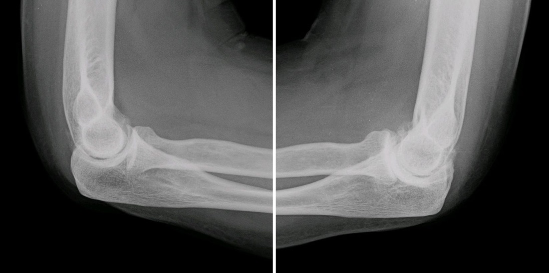
Elbow arthroscopy is a minimally invasive operation in which a camera and fine instruments are introduced into the elbow joint through millimetre-sized incisions. This page covers two main uses: clearing damaged articular cartilage and the bony spurs and loose bodies that accompany it (arthroscopic debridement), and arthroscopic treatment of tennis elbow (lateral epicondylitis) that has not responded to non-operative treatment. The major nerves of the arm run very close to the elbow capsule, which makes elbow arthroscopy one of the most technically delicate arthroscopies in orthopaedics. With the right indication and in experienced hands, it offers small incisions, less pain and fast recovery.
Anatomy of the Elbow
The elbow is made up of three joints within a single capsule: the hinge joint between the humerus and the ulna (humeroulnar), the joint between the humerus and the radial head (radiocapitellar), and the joint between the upper ends of the two forearm bones (proximal radioulnar). Together they allow the elbow to bend and straighten and the forearm to rotate.
Three major nerves run around the joint: the radial nerve and its posterior interosseous branch in front and to the outside, the median nerve in front and to the inside, and the ulnar nerve behind and to the inside. These nerves lie only a few millimetres from the joint capsule. The safety of elbow arthroscopy depends on placing the portals correctly in relation to these nerves and on distending the joint with fluid to push them away from the capsule.
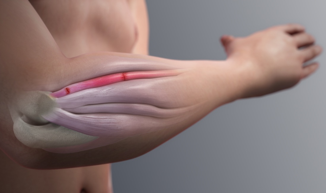
The extensor muscles of the forearm and wrist attach to the bony prominence on the outer side of the elbow (the lateral epicondyle). The origin of the extensor carpi radialis brevis (ECRB) tendon is where the damage of tennis elbow occurs.
Indication 1 — Articular Cartilage Damage and Elbow Arthritis
Cartilage damage and arthritis are less common in the elbow than in the knee and hip, but they occur in heavy manual workers, throwing athletes (baseball, javelin, handball), after elbow fractures and dislocations, and in people who had osteochondritis dissecans at a young age.
- Primary elbow osteoarthritis: Typically in middle-aged men who use their arms heavily; bony spurs (osteophytes) are more prominent than cartilage loss. Osteophytes on the olecranon and coronoid mechanically block full straightening and bending
- Post-traumatic arthritis: Due to joint-surface incongruity after a fracture or dislocation
- Osteochondritis dissecans (OCD) of the capitellum: Particularly in gymnasts and throwers aged 12 - 16; loosening of a cartilage-bone fragment on the capitellum
- Valgus extension overload: Osteophytes and cartilage damage on the back-inner olecranon in throwing athletes
- Loose bodies: Detached cartilage or bone fragments causing locking and catching
- Synovitis and adhesions: From inflammatory disease or after trauma
Indication 2 — Tennis Elbow (Lateral Epicondylitis)
Despite its name, tennis elbow is unrelated to tennis in most patients. It affects about 1 - 3% of the general population, most often between the ages of 35 and 55, in the dominant arm. It is fundamentally not an inflammation but a degenerative tendon disorder (tendinosis) at the ECRB origin caused by repetitive strain: microscopic tears, disorganised collagen and a failed healing response.
The condition is largely self-limiting: about 80 - 90% of patients improve markedly within a year without surgery. Surgery is for the small group whose pain continues to affect daily life and work despite 6 - 12 months of consistent, correctly applied non-operative treatment.
- Repetitive gripping and wrist extension: Using a computer mouse, screwdriver or hammer, carrying heavy bags, working with hand tools
- Racquet sports: Particularly faulty backhand technique, an unsuitable grip size or string tension
- Occupational risk: Butchers, carpenters, painters, plumbers, heavy computer users
- Smoking and obesity: Impair tendon healing and increase the risk
- Diabetes and certain medications (fluoroquinolones): Impair tendon quality
Symptoms
- Cartilage damage and arthritis: Pain at the end of the range when fully straightening or bending the elbow (end-range pain)
- Loss of movement: Particularly inability to straighten the elbow fully; patients often do not notice a few degrees of loss until it progresses and affects daily tasks
- Catching, locking and clicking: With loose bodies
- Morning stiffness and swelling after activity
- Tennis elbow: Pain localised to the outer elbow, just in front of and below the bony prominence
- Tennis elbow: Pain with gripping, shaking hands, lifting a cup, turning a door handle or carrying a bag with the palm down
- Tennis elbow: Reduced grip strength; some patients drop objects
- Pain radiating along the outer forearm
- Ulnar nerve involvement (common with arthritis): Numbness and tingling in the ring and little fingers
Diagnosis
The diagnosis is based on a detailed history, examination and imaging. Occupational and sporting use, previous injuries, previous injections and treatments are reviewed.
Elbow range of movement is measured and compared with the other side, end-range pain is checked, and tender points around the joint are identified. In tennis elbow, pinpoint tenderness just in front of the lateral epicondyle and pain on resisted wrist and middle-finger extension (Cozen's and Maudsley's tests) are typical. Ulnar nerve examination (Tinel's sign, elbow flexion test, hand muscle strength) is performed in every patient; in arthritis it may change the surgical plan.
Differential diagnosis matters: radial tunnel syndrome (compression of the posterior interosseous nerve), referred pain from the neck and lateral ligament instability of the elbow (posterolateral rotatory instability) can mimic tennis elbow and must not be missed.
Plain radiographs (AP and lateral) show osteophytes, loose bodies, joint-space narrowing and OCD lesions; they are usually normal in tennis elbow. CT shows the location and size of osteophytes and loose bodies in detail and is valuable for planning arthritis surgery. MRI assesses cartilage lesions, OCD stability, tendon degeneration and partial tears, and ligament injuries. Ultrasound quickly shows tendon thickening and tears. If there are ulnar nerve symptoms, nerve conduction studies (EMG) are requested.
Non-operative Treatment
In both conditions, treatment starts non-operatively. In tennis elbow this alone is sufficient for the great majority of patients.
- Activity modification: Temporarily reducing the gripping and lifting that provoke pain; learning to lift with the palm facing up
- Physiotherapy and exercise: The foundation of treatment for tennis elbow. Eccentric and isometric strengthening of the forearm extensors, stretching and grip strength work; the effect usually appears within 6 - 12 weeks
- Counterforce brace: Reduces load on the tendon origin and gives relief during activity
- Wrist splint: Particularly at night, reduces pain in some patients
- Analgesics and anti-inflammatory medication (NSAIDs): Short-term, under medical supervision
- Corticosteroid injection: Gives marked relief in the first weeks; however, studies have shown that in tennis elbow, one-year outcomes after corticosteroid injection are worse than with a wait-and-see approach or physiotherapy, with a higher recurrence rate. Repeated injections can thin the tendon and subcutaneous tissue. It is therefore not the first choice for tennis elbow
- PRP (platelet-rich plasma) injection: Used as an option in chronic cases; study results on its effectiveness are inconsistent
- Extracorporeal shock wave therapy (ESWT): An option for resistant cases
- Ergonomic adjustment: Workstation, mouse and keyboard position, tool handle thickness, racquet choice and technique
- Arthritis: Intra-articular injections (corticosteroid, hyaluronic acid), activity modification and range-of-movement exercises; mechanical locking and marked loss of movement do not improve with non-operative treatment
Surgical Treatment — Who Is a Candidate?
- Repeated locking or catching from a loose body: Surgery is appropriate without waiting for non-operative treatment
- Elbow arthritis with loss of movement and end-range pain that affects daily life and work
- Unstable or displaced capitellar OCD lesions
- Olecranon osteophytes from valgus extension overload in throwing athletes
- Tennis elbow: Pain that continues to affect daily life and work despite at least 6 - 12 months of consistent physiotherapy, activity modification and appropriate injections
- Additional advantage of arthroscopy in tennis elbow: The joint can be inspected in the same session, and associated problems (synovial plica, cartilage damage, loose bodies) found and treated
- Not suitable: Tennis elbow in which non-operative treatment was abandoned early; an infected or severely unstable elbow; advanced widespread arthritis (arthroscopy offers limited benefit)
Surgical Options
- Arthroscopic debridement: Removal of damaged, loose cartilage and inflamed synovial tissue
- Loose body removal: The elbow arthroscopy indication with the most predictable result
- Arthroscopic osteocapsular arthroplasty: Shaving of osteophytes on the olecranon and coronoid and release of the contracted anterior capsule; markedly increases range of movement
- OCD treatment: Fixation of stable fragments; removal of unstable fragments with microfracture of the base; mosaicplasty for large lesions
- Arthroscopic ECRB release (tennis elbow): Degenerate tendon tissue is freed from its origin and debrided
- Open (Nirschl) tennis elbow surgery: Removal of degenerate tissue through a small incision; similar success to the arthroscopic method has been reported
- Ulnar nerve release or anterior transposition: Planned in the same session if there is nerve involvement or if preoperative loss of movement is marked (the nerve may be stretched when movement is restored)
Expected Outcomes
Results are most predictable after loose body removal; locking symptoms largely disappear. In elbow arthritis, most patients have less end-range pain and a marked gain in range of movement after arthroscopic debridement and osteocapsular arthroplasty; however, not all lost movement is always regained, and because arthritis is progressive, osteophytes may recur over the years.
After arthroscopic release for resistant tennis elbow, published series report good or excellent results in about 85 - 95% of patients. Complete resolution of pain may take months; full recovery takes 3 - 6 months. The most important factor for success is correct patient selection and excluding conditions that cause similar pain, such as radial tunnel syndrome, before surgery.
Surgery & First 3 Weeks
Elbow arthroscopy is performed under general anaesthesia, often with an additional regional nerve block (supraclavicular or axillary). The block makes postoperative pain control easier; some surgeons delay it until the end of the operation so that a nerve examination can be done immediately afterwards. Most cases are performed as day surgery.
General Information About the Operation
Radiographs and, if needed, CT and MRI are reviewed; the locations of osteophytes and loose bodies are planned in advance. The position of the ulnar nerve is checked on examination: in some people the nerve slips in front of the bony prominence when the elbow bends (subluxation), which creates a risk of injury when the medial portal is made and must be known beforehand. Prophylactic antibiotics are given.
- Duration: Loose body removal or tennis elbow release 45 - 75 minutes; osteocapsular arthroplasty 90 - 150 minutes
- Anaesthesia: General anaesthesia, with a regional block if needed
- Hospital stay: Usually day case or one night
- Position: Lateral decubitus or prone; the arm rests on a support with the elbow hanging at 90°. This lets the anterior nerves fall away from the joint by gravity
- A tourniquet is used
Nerve Safety and Portals
The core safety principle of elbow arthroscopy is to distend the joint with fluid before making the portals. Injecting 20 - 30 ml of fluid stretches the capsule and pushes the anterior nerves a few millimetres away from the joint. Only the skin is incised; deeper tissues are passed with blunt instruments.
- Soft-spot (direct lateral) portal: In the centre of the triangle formed by the radial head, lateral epicondyle and olecranon; the joint is distended through here
- Proximal anteromedial portal: Camera for the anterior compartment; kept in front of the intermuscular septum because of the nearby median nerve and medial antebrachial cutaneous nerve. The position of the ulnar nerve must be confirmed before this portal
- Proximal anterolateral portal: Working portal in the anterior compartment; made about 2 cm above and 1 cm in front of the epicondyle to keep a safe distance from the radial nerve
- Posterolateral and direct posterior (transtriceps) portals: For the olecranon fossa and olecranon tip in the posterior compartment
- Retractors: Blunt retractors through a separate portal lift the capsule to protect the nerves while working on the anterior capsule
Articular Cartilage Debridement and Loose Body Removal
- The anterior compartment is inspected: cartilage surfaces of the coronoid, radial head, capitellum and trochlea, and the anterior capsule
- Loose bodies are removed with a grasper; because they may hide in capsular folds, a systematic search is made
- Loose cartilage flaps and inflamed synovial tissue are removed with a shaver
- Osteophytes on the coronoid tip and in the coronoid fossa are removed with a burr; this removes the mechanical block to full flexion
- The posterior compartment is entered: osteophytes on the olecranon tip and in the olecranon fossa are removed; this allows full extension
- If the anterior capsule is contracted, it is released in a controlled way (capsulectomy); this is the step done with the greatest care because the radial and median nerves lie just in front of the capsule
- If a capitellar OCD is present, its stability is assessed; unstable fragments are removed and microfracture applied to the base, suitable fragments are fixed
- Range of movement is tested on the table; flexion and extension angles are recorded and compared with the preoperative values
Arthroscopic Tennis Elbow (ECRB) Release
- A camera is introduced into the anterior compartment through the proximal anteromedial portal; the lateral capsule and the undersurface of the ECRB tendon are seen
- The joint is inspected: if there is a synovial plica, cartilage damage or a loose body in the radiocapitellar joint, it is treated in the same session
- The lateral capsule is opened, exposing the degenerate, discoloured ECRB tendon tissue behind it
- The degenerate tissue is released from its origin on the lateral epicondyle and debrided with a shaver and radiofrequency device; the bone surface may be lightly freshened to improve blood supply
- Critical boundary: The release is not extended behind the midline of the radial head. The lateral collateral ligament complex (particularly the lateral ulnar collateral ligament) lies just behind this line; damaging it causes elbow instability
- The healthy, superficial part of the extensor muscles (the extensor aponeurosis) is preserved
- Advantages over open surgery: Treatment of intra-articular problems in the same session, a smaller incision and usually an earlier return to work
Discharge Information
Patients usually go home the same day or the next day. At the end of the operation and before discharge, sensation and movement of the hand and fingers are checked; this examination is an important routine after elbow arthroscopy.
After cartilage debridement and osteocapsular arthroplasty the elbow is not immobilised; movement exercises start on the day of surgery or the next day to keep the range gained. A sling is used only for comfort in the first days. A night splint (elbow straight or bent) may be recommended for some patients after capsular release.
After tennis elbow release, a sling or light bandage is used for comfort for the first 1 - 2 weeks; finger, wrist and elbow movements start early, but gripping and lifting are limited.
Ice for 15 - 20 minutes three to four times a day and elevation of the arm above heart level are recommended. Analgesics, anti-inflammatory medication and stomach protection are prescribed. After osteocapsular arthroplasty, some surgeons recommend several weeks of anti-inflammatory medication to prevent heterotopic ossification. Stitches are removed at 10 - 14 days.
Early Postoperative Period (First 3 Weeks)
The priorities in this period depend on the procedure. After cartilage debridement and movement-restoring surgery there is one goal: not losing the movement gained on the table. The elbow is one of the joints most prone to stiffness. After tennis elbow surgery, the priority is to protect the healing tendon origin from early, heavy gripping load.
- Movement exercises (debridement and osteocapsular arthroplasty): 4 - 6 times a day, active and active-assisted work towards full flexion and full extension; forearm rotation is also worked
- Movement exercises (tennis elbow): Finger, wrist and elbow movements within the pain limit; no heavy gripping, lifting or resisted wrist extension for the first 4 - 6 weeks
- Swelling control: Elevation of the arm above heart level, ice and finger pumping exercises
- Wound care: Portal sites kept dry for 10 - 14 days; elbow portals may leak joint fluid for a few days — report increased or cloudy discharge
- Nerve monitoring: Numbness in the ring and little fingers (ulnar nerve), numbness in the thumb and index finger (median nerve), or inability to lift the wrist and fingers (radial nerve) must be reported immediately
- Daily life: Using the arm for light tasks (eating, writing, personal care) is encouraged; it helps both range of movement and muscle activation
Follow-up
- Week 1: Range-of-movement measurement, nerve examination and review of the exercise programme
- Days 10 - 14: Removal of stitches
- Week 6: Range-of-movement and strength assessment; decision to start strengthening for tennis elbow
- Month 3 and month 6 reviews
Risks & Complications
Elbow arthroscopy is safe in experienced hands and serious complications are rare. However, the closeness of the nerves around the joint to the capsule makes this arthroscopy more delicate than knee or shoulder arthroscopy. The most important risk is nerve injury; most nerve problems are temporary.
Risks Specific to Non-operative Treatment
- Long course in tennis elbow: Recovery with non-operative treatment may take 6 - 12 months, affecting work and daily life
- Repeated corticosteroid injections: Thinning of subcutaneous fat, skin lightening, weakening of tendon tissue and a higher long-term recurrence rate
- Arthritis: Mechanical locking and loss of movement do not improve with non-operative treatment; loss of movement may progress as osteophytes grow
- Loose bodies left in the joint: Repeated locking may cause further cartilage damage
Surgical Risks — Common
- Temporary nerve problems: A temporary change in sensation or weakness after elbow arthroscopy is reported in about 1 - 3% of cases in studies; most resolve on their own within weeks or months
- Prolonged leakage of joint fluid from a portal
- Superficial portal infection (usually treated with antibiotics)
- Postoperative pain, swelling and bruising
- Temporary elbow stiffness — improves with regular exercise
- Small areas of numbness from injury to superficial sensory branches at the portal sites
Surgical Risks — Less Common
- Permanent nerve injury: Ulnar, radial (particularly the posterior interosseous branch) or median nerve; rare but serious. The risk is higher in patients undergoing capsular release and osteocapsular arthroplasty
- Ulnar nerve irritation: When range of movement is markedly increased, particularly flexion, the nerve may be stretched; this is why nerve release is planned in the same session in patients with very limited preoperative flexion
- Loss of the movement gained: The elbow re-stiffens quickly when the exercise programme is not followed
- Heterotopic ossification: Bone formation in soft tissue; may restrict movement
- Deep joint infection (under 1%): Requires arthroscopic washout and antibiotics
- Specific to tennis elbow surgery: Damage to the lateral ligament from extending the release too far back, causing elbow instability (posterolateral rotatory instability) — rare but difficult to treat
- Specific to tennis elbow surgery: Persistent pain, which may reflect a coexisting condition such as radial tunnel syndrome
- Complex regional pain syndrome (CRPS): Rare
- Instrument breakage or retained fragments: Very rare
Risk Factors
- Previous elbow surgery or ulnar nerve transposition: Normal anatomy is altered, making portal placement riskier
- Ulnar nerve subluxation (slipping in front of the bony prominence): If not identified beforehand, risk of injury when the medial portal is made
- A severely stiff elbow: Reduced joint volume means the nerves cannot be pushed far enough from the capsule
- Inflammatory diseases such as rheumatoid arthritis that thin the capsule
- Smoking: Impairs tendon and wound healing
- Poorly controlled diabetes
- Poor adherence to the exercise programme: The most common cause of recurrent stiffness after debridement
- Tennis elbow: Early return to work or sport with heavy loads
Contact Your Doctor Immediately If
- You cannot lift your wrist or fingers
- Numbness, tingling or weakness in the fingers is progressing
- Pain or swelling suddenly increases
- You have a temperature above 38 °C
- There is cloudy or foul-smelling discharge from a portal, or redness and warmth around the wound
- Your fingers become blue, cold or pale
- The movement gained after surgery is rapidly being lost
Long-term Outcomes
After loose body removal and arthroscopic debridement, most patients achieve a pain-free, more mobile elbow. In elbow arthritis, osteophytes may re-form over time, so partial recurrence of symptoms over the years is possible; if advanced arthritis develops, options such as elbow replacement are considered, although this is uncommon.
After arthroscopic release for resistant tennis elbow, good results are largely maintained in the long term and the recurrence rate is low. Return to desk work usually takes 1 - 3 weeks and to heavy manual work 6 - 12 weeks. Full recovery of grip strength may take 3 - 6 months. Ergonomic adjustment and maintaining forearm muscle strength are the most effective ways to prevent recurrence.
Rehabilitation
Rehabilitation after elbow arthroscopy follows two different paths depending on the procedure. After cartilage debridement and movement-restoring surgery, the core rule is early and frequent movement; the elbow is one of the joints that stiffens fastest when left still. After tennis elbow surgery, movement starts early but load comes late; the healing tendon origin is protected from heavy gripping for weeks.
After Cartilage Debridement and Osteocapsular Arthroplasty
- Week 0 - 1: Movement exercises start on the day of surgery or the next day. Full flexion and full extension work plus forearm rotation 4 - 6 times a day. Sling only briefly, for comfort. Ice and elevation. Night splint if needed.
- Weeks 1 - 3: Range of movement is measured at every visit; the aim is to keep the range gained on the table. Supervised physiotherapy 2 - 3 times a week. The arm is used for daily tasks.
- Weeks 3 - 6: Light strengthening begins while movement work continues: forearm, biceps, triceps and grip work with light resistance.
- Weeks 6 - 12: Strengthening intensifies. Full return to daily and work activities. Stretching continues.
- Months 3 - 6: Gradual return to sport and heavy work. A graded throwing programme for throwing athletes.
After Arthroscopic Tennis Elbow Release
- Weeks 0 - 2: Sling or light bandage for comfort. Finger, wrist and elbow movements within the pain limit. No heavy gripping or lifting. Ice and elevation.
- Weeks 2 - 6: Full range of movement is the goal. Gentle stretching of the wrist extensors. Light, pain-free isometric work. Return to desk work is usually possible in this period.
- Weeks 6 - 12: Graded strengthening: eccentric exercises for the wrist extensors, grip strength work, forearm rotation strengthening. Resistance is increased using pain as a guide.
- Months 3 - 4: Gradual return to heavy manual work. Gradual return to racquet sports together with a technique review.
- Months 4 - 6: Return to full strength. Grip strength usually approaches the other side in this period.
Key Exercises
- Elbow flexion-extension: With help from the good arm, holding for 20 - 30 seconds at the end of the movement
- Forearm rotation: Elbow bent to 90° and close to the body, turning the palm up and down; a light hammer held further along the handle can increase difficulty
- Wrist extensor stretch: Elbow straight, palm down, the other hand gently bends the wrist downwards
- Eccentric wrist extension (tennis elbow): A light weight is lifted with help from the other hand and lowered slowly (over 3 - 5 seconds) with the operated hand. Mild discomfort is acceptable; sharp pain is not
- Grip exercises: With a soft ball or grip device, with gradually increasing resistance
- Movement rather than rest: Using the arm for daily tasks is a natural continuation of the exercise programme
Return-to-sport and Work Criteria
- Pain-free elbow range of movement close to the other side
- Grip strength at least 85 - 90% of the other side
- Symmetrical wrist extension and forearm rotation strength
- Pain-free completion of work- or sport-specific movements
- No pain or swelling after activity
- Uneventful completion of a graded throwing programme in throwing athletes
- Approval from the surgeon and physiotherapist
Recommended and Avoided Activities
- Recommended: Light daily tasks from early on, walking, lower-body conditioning
- Recommended: Swimming (once healed; from weeks 3 - 4 in debridement patients)
- Tennis elbow — avoid in the first 6 weeks: Heavy gripping, lifting with the palm down, using a hammer or screwdriver, carrying heavy bags
- Avoid in the first 3 months: Racquet sports, gym exercises that strain the wrist and elbow
- Debridement patients — avoid: Keeping the elbow in the same position without moving for long periods
- Throwing athletes: Uncontrolled return to throwing without a graded programme
Lifestyle and Recovery Advice
- Do your exercise programme every day; after debridement, regular movement is the only remedy for elbow stiffness
- Lift with your palm facing up; this reduces the load on the tendon origin at the outer elbow
- Review your work environment: mouse and keyboard position, tool handle thickness, breaks from repetitive movements
- If you play racquet sports, have your grip size, string tension and backhand technique assessed by a coach
- Do not smoke; it markedly impairs tendon and tissue healing
- If you have diabetes, keep your blood glucose tightly controlled
- Maintain forearm muscle strength in the long term; strong muscles protect the tendon origin
- Be patient: Complete resolution of pain after tennis elbow surgery may take 3 - 6 months
- Attend regular follow-up (week 1, week 2, week 6, month 3)
What to Expect
After loose body removal and cartilage debridement, return to daily activities usually takes 1 - 3 weeks and return to sport 6 - 12 weeks. After osteocapsular arthroplasty, the gain in range of movement is marked in the first weeks; whether it lasts depends on adherence to the exercise programme. Full recovery takes 3 - 6 months.
After tennis elbow release, return to desk work takes 1 - 3 weeks and to heavy manual work 6 - 12 weeks. Complete resolution of pain and recovery of grip strength may take 3 - 6 months; mild pain continuing in the first weeks is expected. Tenderness and small patches of numbness at the portal sites settle within a few months.
Is This Treatment Right for You?
Every patient's condition is different. After a detailed examination and imaging, the treatment plan that suits you best will be prepared.
Would you like more information about Elbow Arthroscopy?
Book Appointment