Talar Osteochondral Defect
Arthroscopic and open surgical treatment of talar osteochondral defects
Overview
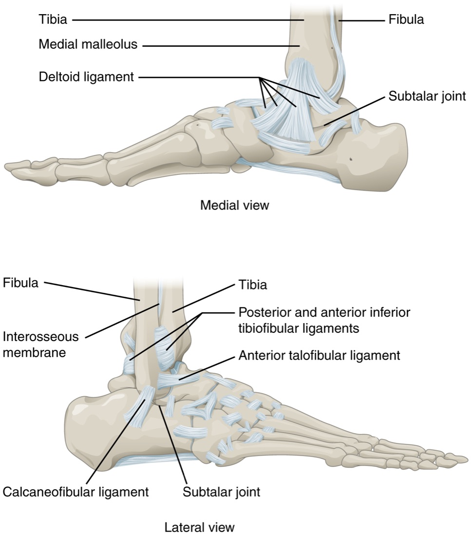
An osteochondral defect (osteochondral lesion / OCL) of the talus is damage to the cartilage of the talar dome and the subchondral bone beneath it. It may take the form of a small crack in the joint surface, a loosened fragment, or complete separation of the cartilage-bone complex (a "flap" or a loose body). It commonly develops after a single or repeated ankle sprain; 70–90% of cases are traumatic in origin. Left untreated it can lead to progressive loss of articular cartilage and ankle arthritis.
Anatomy of the Ankle Joint and the Talus
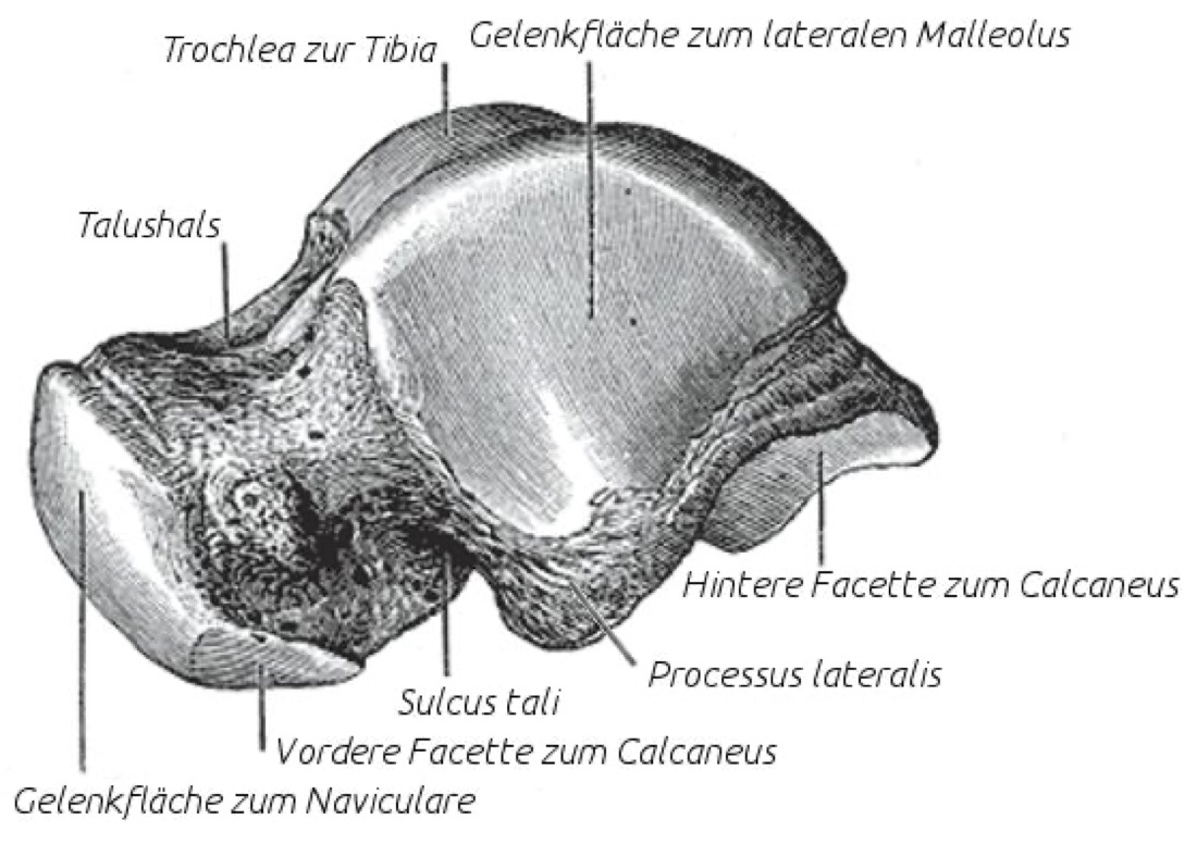
The ankle is a hinge joint formed by three bones: the shin bone (tibia), the calf bone (fibula) and the talus. The talus lies between the tibia and the calcaneus and is a unique bone with no muscle attachments; approximately 60% of its surface is covered with cartilage and its blood supply is limited.
Because of these features, damaged talar cartilage has a poor capacity to heal and, if not treated correctly, may progress to advanced ankle arthritis.
Location of the Lesion
The commonest locations are the posterior part of the medial talar dome and the anterior-central part of the lateral talar dome; medial lesions are generally deeper and more cystic, while lateral lesions are more superficial and traumatic. Less commonly the cause is idiopathic or genetic (juvenile osteochondritis dissecans).
Berndt–Harty Classification (Plain Radiographs)
- Stage I: a small compression fracture beneath the cartilage (not visible or only faintly visible radiographically)
- Stage II: a partially detached cartilage-bone fragment (still attached in place)
- Stage III: a completely detached but non-displaced (in situ) fragment
- Stage IV: a displaced free fragment (loose body)
Hepple (MRI) Classification
- Stage I: articular cartilage damage only
- Stage II: cartilage damage plus minimal fluid or oedema in the subchondral bone
- Stage III: cartilage damage plus a detached fragment (in place)
- Stage IV: cartilage damage plus a detached and displaced fragment
- Stage V: subchondral cystic lesion
Classification by Lesion Size
- Small: <1 cm² (<10 mm diameter)
- Medium: 1–1.5 cm²
- Large: >1.5 cm²
- Lesion size is one of the most important factors in choosing the surgical technique
Symptoms
- Chronic ankle pain: a deep pain that increases with activity and partly settles with rest
- Swelling: oedema and joint effusion, particularly after activity
- Clicking or catching: particularly with stage III–IV lesions
- Sudden locking: in the presence of a loose body
- Giving way: particularly where there is associated lateral instability
- Restricted movement: particularly in dorsiflexion and plantarflexion
- Morning stiffness: consistent with cartilage damage
- A history of previous ankle sprain: present in most patients
Diagnosis
Diagnosis is based on a detailed history, physical examination and imaging. The history of injury (particularly the number of previous sprains), the level of sporting activity and comorbidities are reviewed. Examination assesses swelling, points of tenderness (particularly along the joint line), range of motion, stability tests (anterior drawer, talar tilt) and gait.
Plain radiographs are the first-line investigation; standing AP, mortise and lateral views are standard. Early-stage lesions may not be visible on radiographs. CT gives detailed information on bone anatomy and cystic lesions. MRI is the gold standard for diagnosing a talar OCD; it demonstrates the cartilage damage, subchondral bone oedema, lesion size, the stability of the fragment and any associated pathology. In selected cases diagnostic arthroscopy provides definitive diagnosis and treatment in the same sitting.
Non-operative (Conservative) Treatment
This is tried in acute, non-displaced lesions (particularly in children and adolescents), in patients at surgical risk, and in adults with mild symptoms.
- Activity modification and weight control
- Non-weight bearing or partial weight bearing for 4–6 weeks (cast, walker boot)
- Analgesics and anti-inflammatory medication (NSAIDs)
- Physiotherapy: range of motion, proprioception and muscle strengthening
- Intra-articular injections (corticosteroid, hyaluronic acid, PRP)
- Success rate: 50–60% in children and adolescents, 25–50% in adults
Surgical Treatment — General Approach
Surgery is planned in cases that do not respond to non-operative treatment; in displaced stage III–IV lesions; in large (>1.5 cm²) and cystic lesions; and in active sporting patients. The technique is chosen according to the size and depth of the lesion:
- Small lesions (<1–1.5 cm²): bone marrow stimulation (microfracture / drilling) — arthroscopic
- Stable, in situ stage II–III lesions: fragment fixation (bioabsorbable screw or pin)
- Lesions with a subchondral cyst: retrograde drilling plus bone graft
- Medium to large lesions (>1.5 cm²): mosaicplasty / OAT (autologous osteochondral graft) — open (a medial malleolar osteotomy is often required)
- Large or cystic lesions and salvage procedures: osteochondral allograft transplantation — open
- Cell- and matrix-based techniques: ACI / MACI (autologous chondrocyte implantation)
- Matrix-assisted regeneration: AMIC (collagen/hyaluronic membrane plus microfracture)
- Removal of a loose body — arthroscopic (always combined with additional treatment)
- Salvage surgery: ankle arthrodesis — open
Advantages
The principal advantage is the repair or reconstruction of the cartilage surface. This provides pain control, reduces the risk of developing advanced ankle arthritis and preserves functional range of motion.
- Arthroscopic techniques are minimally invasive: small incisions, less pain, a lower risk of infection and faster recovery
- Mosaicplasty / OAT transfers true hyaline cartilage; long-term results in large lesions are superior to microfracture
- Allograft transplantation allows reconstruction of the joint surface in very large lesions and offers an alternative to arthrodesis or arthroplasty
- Fixation of stable fragments preserves the hyaline cartilage
- Operative time: arthroscopic microfracture 30–60 minutes; mosaicplasty 90–150 minutes; allograft / ACI 120–180 minutes
- Most arthroscopic cases are day cases; a stay of 1–2 days is usual after major open surgery
Disadvantages
- There are surgery-specific risks: wound infection, deep infection, nerve injury (particularly the superficial peroneal, sural and saphenous nerves), implant irritation and, rarely, vascular injury
- After mosaicplasty, temporary pain, swelling or patellofemoral symptoms may develop at the donor site (the knee on the same side)
- A medial malleolar osteotomy is usually required for medial talar dome lesions; implant irritation, rarely non-union or an intra-articular screw problem may occur at the osteotomy site
- The weight-bearing protocol is long: non-weight bearing is required for 4–6 weeks after microfracture and 6–8 weeks after mosaicplasty, and longer after allograft or ACI
- The fibrocartilage produced by microfracture is biomechanically weaker than hyaline cartilage; it may wear over time and symptoms may return
- Allograft transplantation is limited by tissue availability and carries a risk that the graft will not integrate
Why Surgery for a Talar OCD?
Because the blood supply to the talus is limited, OCD lesions cannot heal completely on their own. Untreated, the lesion may enlarge, undergo cystic change and cause collapse of the joint surface; in the long term this progresses to advanced post-traumatic ankle arthritis and creates the need for arthrodesis or arthroplasty.
With early intervention, fibrocartilage repair is possible in small lesions and hyaline cartilage reconstruction in medium to large lesions, and long-term patient satisfaction is high.
Who is a Candidate?
Surgery for a talar OCD is performed for lesions that have not responded to at least 3–6 months of non-operative treatment, or that are markedly displaced (stage III–IV) from the outset.
- Patients with chronic ankle pain that increases with activity
- Individuals with a stage II–V lesion on MRI
- Patients with episodes of locking due to an intra-articular loose body
- Lesions showing subchondral cystic change
- Cases being considered for salvage surgery after failed non-operative treatment or failed initial surgery
- Patients whose general health is suitable for general anaesthesia and surgery
Expected Outcomes
With the correct indication and compliant rehabilitation, patient satisfaction after talar OCD surgery is 75–90%. In small lesions treated with microfracture, short- to medium-term results (2–5 years) are excellent; however, in the longer term (>5 years) symptoms may recur as the fibrocartilage wears.
The rate of good to excellent results at 5–10 years after mosaicplasty / OAT is 85–90%. Allograft transplantation is promising in selected large lesions as an alternative to arthrodesis or arthroplasty. Return to sport may take 4–6 months after arthroscopic microfracture, 6–9 months after mosaicplasty and 9–12 months after allograft or ACI.
Surgery & First 3 Weeks
Surgery for a talar OCD is performed under regional anaesthesia (a popliteal block and/or saphenous nerve block), spinal/epidural anaesthesia or general anaesthesia. A regional block is effective for pain control in the first 12–24 hours after surgery. Arthroscopic cases are generally performed as day cases; a stay of 1–2 days may be required for open surgery (particularly with a medial malleolar osteotomy) and for major reconstructions.
General Information About the Operation
- Duration: arthroscopic microfracture / debridement 30–60 minutes; mosaicplasty (with a medial malleolar osteotomy) 90–120 minutes; allograft transplantation or ACI 90–120 minutes
- Anaesthesia: regional block, spinal/epidural or general anaesthesia
- Hospital stay: 1 day for arthroscopic cases; 1–2 days for open and complex cases
- Radiographs, MRI and, where required, CT are reviewed for preoperative planning; prophylactic antibiotics are given
Surgical Approaches
- Arthroscopic anterior approach: anteromedial and anterolateral portals; visualisation of the anterior cartilage surface
- Arthroscopic posterior approach: posteromedial and posterolateral portals; particularly for posterior medial talar dome lesions
- Medial malleolar osteotomy (open): the most commonly used open approach for access to the posterior part of the medial talar dome; the osteotomy is fixed back with screws
- Lateral malleolar osteotomy / temporary removal of the fibula: for the lateral talar dome (rarely)
- Anterior arthrotomy: for some anterior lesions and for allograft placement
- Hybrid approaches: arthroscopic assessment plus a limited open procedure
Arthroscopic Debridement and Removal of Loose Bodies
- This is the first step in every case. Free cartilage or bone fragments within the joint (loose bodies) are removed with a grasper or small instruments
- Unstable or unattached cartilage fragments are cleared
- A partial synovectomy is added where there is associated synovitis
Microfracture / Drilling / Nanofracture
This is the most commonly used bone marrow stimulation technique. Indication: small lesions (<1–1.5 cm²) with intact subchondral bone support.
- The edges of the lesion are debrided back to stable hyaline cartilage (creating vertical walls)
- Fine holes (1–2 mm) are made in the subchondral bone with an awl (microfracture), a K-wire (drilling) or dedicated nanofracture instruments, approximately 3–4 mm apart
- The mesenchymal stem cells in the blood that seeps from these holes lead to the formation of fibrocartilage
- Advantages: minimally invasive, no additional implants required, short operative time
Retrograde Drilling
- Preferred in stable lesions where the overlying cartilage is intact and there is only oedema or a small cyst in the subchondral bone
- A tunnel is drilled towards the lesion under fluoroscopic guidance from the lateral or medial surface of the talus, through the sinus tarsi or by a transmalleolar approach
- The tunnel may be filled with bone graft or a substitute
- Advantage: healing is stimulated without breaching the intact cartilage surface
In Situ Fragment Fixation
- Stage II–III stable but detached cartilage-bone fragments are repositioned and fixed
- Bioabsorbable screws or pins, small metal screws or headless compression screws are used
- This has the advantage of preserving hyaline cartilage
- Indication: a young patient, an acute displaced fragment, and good fragment quality
AMIC (Matrix-Assisted Microfracture)
- A collagen or hyaluronic membrane is placed over the lesion after microfracture to retain the mesenchymal stem cells at the site
- This is a single-stage method that does not require cell culture
- It may be preferred in medium-sized lesions (1–3 cm²) where microfracture alone is insufficient
- It can be performed arthroscopically or open, and is more practical than ACI
Mosaicplasty / OAT (Osteochondral Autograft Transfer)
True hyaline cartilage and subchondral bone are transferred together as a unit. Indication: medium to large lesions (1–3 cm²), particularly where subchondral bone support is compromised.
- Donor site: one or more cylindrical bone-cartilage grafts 4–10 mm in diameter are harvested from the knee on the same side (the non-weight-bearing part of the medial femoral condyle or the trochlear ridge)
- Recipient site: cylindrical tunnels of the same diameter are drilled into the base of the lesion in the talus
- The grafts are inserted into the tunnels by press-fit; the cartilage surfaces are set flush with the surrounding intact cartilage
- A medial malleolar osteotomy is usually required for medial talar dome lesions; it is fixed back with two or three cannulated screws at the end of the operation
- Advantage: transfer of hyaline cartilage, biomechanically superior to fibrocartilage
Osteochondral Allograft Transplantation (OCA)
- The lesion is reconstructed with a fresh talar or femoral allograft from a donor
- It is usually inserted through an anterior arthrotomy; a medial or lateral malleolar osteotomy may be required
- Indication: large (>3 cm²) and deep cystic lesions; failed previous treatment; an alternative to arthrodesis or arthroplasty in young patients
- Advantage: near-complete reconstruction of the joint surface is possible in very large lesions
- Disadvantages: limited tissue availability, slow allograft integration, and a (low) risk of immune response
Autologous Chondrocyte Implantation (ACI / MACI)
This is a two-stage (classic ACI) or single-stage (matrix-based MACI) cellular treatment. Indication: medium to large lesions (>2 cm²), particularly after failed microfracture.
- Stage 1: a small biopsy is taken arthroscopically from a non-weight-bearing area of cartilage; the cells are cultured in the laboratory for 4–6 weeks
- Stage 2: the lesion is prepared through an open procedure; the cultured chondrocytes are placed beneath a periosteal flap (classic ACI) or a collagen membrane (MACI)
- Advantage: formation of hyaline-like cartilage; long-term results are promising
Open ORIF and Bone Grafting
- Open ORIF (in situ fixation): for large, acute, displaced stage III–IV fragments. The lesion is reached through a medial malleolar osteotomy or an anterior arthrotomy; the bed beneath the fragment is debrided, bone graft is added and the fragment is fixed with cannulated or bioabsorbable screws
- Open bone grafting (cystic lesions): for large subchondral cysts the cyst is curetted and filled with autologous (iliac crest) or allogeneic bone graft or a bone substitute. The overlying cartilage surface is preserved or reconstructed
Ankle Arthrodesis (Salvage Surgery)
- Performed for advanced arthritis that develops after several failed OCD treatments
- The joint surfaces are removed and the tibia is fused to the talus (with screws or a plate)
- Joint movement is permanently lost; however, pain is reduced dramatically
Implant Options
- Bioabsorbable screws / pins — for in situ fragment fixation
- Headless compression screws (Herbert, Acutrak) — countersunk beneath the cartilage surface
- Cannulated screws — to fix a medial malleolar osteotomy (3.5–4.5 mm)
- Collagen / hyaluronic membranes — for AMIC and MACI
- Autologous bone graft (iliac crest, distal tibia) — for cystic lesions
- Allogeneic bone graft or a synthetic bone substitute — for cystic lesions
- Mosaicplasty instrumentation (osteochondral transfer systems)
Discharge Information and Weight-Bearing Protocol
Arthroscopic cases are usually discharged the same day; a stay of 1–2 days may be required after open surgery or a medial malleolar osteotomy. After surgery the ankle is generally immobilised in a short leg cast, a walker boot or a volar splint. The weight-bearing protocol varies according to the procedure performed:
- Arthroscopic debridement / removal of a loose body: weight bearing as tolerated (in a walker boot)
- Microfracture / nanofracture: non-weight bearing for 4–6 weeks, then graduated
- Retrograde drilling: non-weight bearing for 4–6 weeks (this may be shorter if the cartilage surface is intact)
- In situ fragment fixation: non-weight bearing for 6–8 weeks
- Mosaicplasty / OAT: non-weight bearing for 6–8 weeks
- Allograft transplantation: non-weight bearing for 8–12 weeks
- ACI / MACI: non-weight bearing for 6–8 weeks
- Medial malleolar osteotomy: the osteotomy healing protocol is followed (usually 6 weeks non-weight bearing)
- Ice for 15–20 minutes three to four times a day and elevation are recommended. Analgesics, anti-inflammatories, gastric protection and anticoagulation (until weight bearing) are prescribed. Sutures are removed on day 10–14
Early Postoperative Period (First 3 Weeks)
The aims during this period are wound healing, control of swelling, pain management, maintaining movement of the toes and of the hip and knee, minimising muscle atrophy and continuing thromboprophylaxis.
- Wound monitoring: discharge, marked redness, increased warmth or wound edge separation are monitored. It is important not to wet the wound for the first 10–14 days
- Control of swelling: keeping the leg above the level of the heart (elevation), ice and compression stockings reduce oedema
- Weight-bearing protocol: strict compliance with the programme set for the procedure performed is critical. Early weight bearing increases the risk of graft failure and revision
- Early passive movement: after microfracture, mosaicplasty, ACI/MACI and AMIC, early passive and active-assisted ankle movement exercises support cartilage healing. A CPM machine or manual passive exercises are recommended
- Toe, hip and knee movement: performed three to four times a day within the splint or boot; this reduces swelling and prevents stiffness in the more distant joints
- Pain: once the regional block has worn off, prescribed medication, ice and elevation control the pain
- Thromboprophylaxis: the risk increases during the non-weight-bearing period. Anticoagulation, compression stockings, toe movement and hip and knee exercises are used together
- Nerve monitoring: the superficial peroneal, sural and saphenous nerves lie close to the portals and open incisions; report any numbness or tingling in the toes
Follow-up
- Days 10–14: suture removal and check radiograph
- Weeks 4–6: decision on weight bearing and, where required, a check radiograph of the osteotomy
- Month 3: rehabilitation review and, where required, MRI
- Month 6 and one-year review
Risks & Complications
Surgery for a talar OCD is one of the minimally invasive areas of modern orthopaedics; arthroscopic techniques in particular are safe and the rate of serious complications is low. Nevertheless every operation carries certain risks, and the risk profile varies with the size of the lesion and the procedure performed.
Common (Under 5%)
- Superficial wound infection (usually treated with antibiotics)
- Postoperative pain, swelling and temporary bruising
- Temporary numbness or tenderness at the portal sites
- Transient symptoms related to anaesthesia such as nausea and dizziness
- Temporary ankle stiffness — this resolves with physiotherapy
- Prolonged swelling (this may continue for 6–12 months)
Less Common
- Deep infection: 1–2%; may require revision, implant removal and prolonged antibiotics
- Nerve injury: the superficial peroneal nerve (anterolateral portal), the sural nerve (posterolateral portal) and the saphenous nerve (anteromedial portal and medial approaches); temporary numbness is common, permanent neuropathy rare
- Vascular injury (very rare)
- Deep vein thrombosis (DVT) and pulmonary embolism (PE): the risk increases during the non-weight-bearing period; largely prevented by anticoagulation
- Implant failure: loosening or breakage of a screw
- Complex regional pain syndrome (CRPS): rarely seen
- Heterotopic ossification (bone formation in soft tissue)
Risks Specific to Microfracture / Drilling and Retrograde Drilling
- The formation of fibrocartilage is the natural result of microfracture; it is biomechanically weaker than hyaline cartilage and may wear over time
- Return of symptoms in the late period (20–30%, over the years)
- Cystic degeneration in the subchondral bone
- Intralesional osteophyte formation (growth of subchondral bone into the base of the lesion)
- With retrograde drilling: inadequate drilling or a misdirected tunnel; damage to the subtalar joint with a sinus tarsi approach (rare)
Risks Specific to In Situ Fragment Fixation
- Failure of the fragment to unite / failure of fixation (10–25%)
- The screw head penetrating into the joint (cartilage damage)
- Foreign body reaction to bioabsorbable implants (rare)
Risks Specific to Mosaicplasty / OAT
- Donor site (knee) morbidity: pain, swelling and patellofemoral symptoms (10–30%); these mostly settle over time
- Failure of the graft to integrate, or plug subsidence
- Level mismatch: the graft surface not matching the recipient cartilage (sitting proud or recessed)
- Gaps between plugs that do not fill with fibrocartilage
Risks Specific to Allograft and ACI / MACI
- Allograft: delayed or failed graft integration; collapse of the graft; immune reaction (rare); risk of disease transmission (very low with modern tissue bank processing); delay in obtaining tissue
- ACI / MACI: periosteal or membrane hypertrophy (more common with classic ACI; may require debridement)
- ACI / MACI: graft delamination (separation of the cartilage from beneath)
- ACI: the requirement for two-stage surgery; contamination during cell culture (very rare)
Risks Specific to a Medial Malleolar Osteotomy
- Non-union at the osteotomy site (1–5%)
- Malunion (the osteotomy healing in the wrong position)
- Implant irritation; some patients may require screw removal
- An intra-articular screw (disruption of the medial mortise)
Long-term Complications
- Return of symptoms (need for revision): particularly within 5 years after microfracture
- Traumatic / post-OCD arthritis: may develop over the years and progress to arthrodesis or arthroplasty
- Enlargement of the lesion (despite surgery)
- Cystic degeneration (particularly after failed microfracture)
- Permanent restriction of movement; chronic pain
Factors that Increase Risk
- Smoking (markedly increases the risk of problems with wound and cartilage healing and of infection)
- Poorly controlled diabetes (impaired wound healing)
- Obesity (additional load on the ankle)
- Age (the capacity for cartilage healing is lower above 40–50)
- Large (>1.5 cm²) and cystic lesions
- Associated ankle instability (if untreated, the OCD recurs)
- Malalignment (a varus deformity with medial lesions)
- Failed previous surgery
Contact Your Doctor Immediately If You Have
- Sudden increase in pain or swelling
- Fever above 38 °C
- Discharge, malodour or separation of the wound
- Increasing numbness, tingling or loss of strength in the foot or toes
- Bruising, coldness or pallor of the toes (suspected vascular problem)
- Firm, one-sided swelling or redness of the leg (suspected DVT)
- Shortness of breath or chest pain (suspected pulmonary embolism)
- Excessive tightness or unbearable pain within the cast or boot
Long-term Outcomes
With the correct indication, an appropriate surgical technique and compliant rehabilitation, the results of talar OCD surgery are generally good. After microfracture, 70–85% of patients are satisfied in the short to medium term; however, in 20–40% symptoms recur in the longer term (>5 years). The rate of good to excellent results at 5–10 years after mosaicplasty / OAT is 85–90%.
Allograft transplantation and ACI/MACI give promising results in selected large lesions. Progression of the lesion and the development of advanced arthritis in the long term remain possible despite treatment; arthrodesis, or ankle arthroplasty in selected patients, then comes into consideration. Stopping smoking, weight control, correction of any associated pathology and disciplined rehabilitation are the fundamental determinants of long-term success.
Rehabilitation
Rehabilitation after talar OCD surgery is the most important part of recovery and directly affects the success of the operation. The aims are to control swelling and pain, support cartilage healing, preserve joint range of motion, restore the strength of the calf and intrinsic foot muscles, and restore proprioception. The general principle is early passive movement with delayed weight bearing: passive movement stimulates cartilage regeneration, while early weight bearing is prohibited because it causes the new cartilage to collapse.
After Arthroscopic Microfracture / Nanofracture
- Weeks 0–2: Non-weight bearing with crutches. A walker boot or short leg cast. Early passive and active-assisted ankle movement exercises (with a CPM machine or manually, 4–6 hours a day). Toe, hip and knee exercises. Ice and elevation.
- Weeks 2–6: Non-weight bearing continues. Active movement exercises (flexion, extension, gentle inversion and eversion). Quadriceps and calf isometric exercises. Stationary cycling (with light resistance).
- Weeks 6–8: Graduated weight bearing (partial → full). Transition from the walker boot to a normal shoe. Calf and intrinsic foot strengthening.
- Months 2–4: Full weight bearing; gait normalises. Strengthening with a resistance band. Balance and proprioception exercises. Swimming.
- Months 4–6: Beginning light running in a straight line (with the surgeon's approval). Graduated introduction of sport-specific drills.
- Months 6–12: Full return to sport and high-impact activity with the surgeon's approval. Maturation of the fibrocartilage takes 12 months.
After In Situ Fragment Fixation
- Weeks 0–6: Non-weight bearing with crutches. A walker boot or cast. Early passive movement exercises. Toe, hip and knee exercises.
- Weeks 6–8: The position of the fragment is confirmed on a check radiograph; graduated weight bearing begins. Active movement exercises.
- Months 2–4: Full weight bearing. Strengthening and proprioception. Swimming and stationary cycling.
- Months 4–9: Graduated return to sport; high-impact activity at months 9–12.
After Mosaicplasty / OAT
- Weeks 0–6/8: Non-weight bearing with crutches. Early passive movement exercises are critical. Rehabilitation of the donor site (the knee) proceeds in parallel (quadriceps strengthening, knee movement).
- Weeks 8–12: Graduated weight bearing. Active movement exercises. Light strengthening. Stationary cycling and swimming.
- Months 3–6: Full weight bearing; gait normalises. Strengthening and proprioception intensify. Beginning light running in a straight line (around months 4–5).
- Months 6–9: Graduated return to sport-specific drills.
- Months 9–12: Full return to contact and high-impact activity with the surgeon's approval.
After Allograft Transplantation
- Weeks 0–8/12: Non-weight bearing (graft integration takes longer). Early passive movement exercises. Quadriceps and hip exercises.
- Weeks 12–16: Graduated weight bearing. Active movement exercises.
- Months 4–9: Strengthening and proprioception. Return to low-impact activity.
- Months 9–12: Graduated return to sport; full high-impact activity at months 12–18.
After ACI / MACI
- Weeks 0–6: Non-weight bearing; a CPM machine is recommended. Early movement exercises are critical for cartilage maturation. Toe and distant joint exercises.
- Weeks 6–12: Graduated weight bearing. Active movement exercises. Light strengthening.
- Months 3–6: Full weight bearing; return to low-impact activity.
- Months 6–12: Graduated return to sport.
- Months 12–18: Complete maturation of the cartilage; full return to high-impact activity.
Special Notes for Cases with a Medial Malleolar Osteotomy
- The osteotomy takes 6 weeks to heal; the weight-bearing protocol is set taking both the OCD and the osteotomy into account
- Screw removal at the osteotomy site is usually not required; if it causes pain, it is considered after months 9–12
- Active inversion (turning the foot inwards) is limited for the first 6 weeks
Criteria for Return to Sport and Activity
- Full, pain-free ankle range of motion
- Calf muscle strength reaching at least 85–90% of the other leg
- At least 25 repetitions on the single-leg heel rise test
- Single-leg balance tests (30 seconds with the eyes closed)
- 90% symmetry on single-leg hop tests
- No swelling or pain after activity
- Pain-free completion of sport-specific drills
- Approval of the surgeon and physiotherapist (with a check MRI where required)
Recommended Activities
- Stationary cycling and the elliptical trainer (non-weight-bearing cardiovascular activity)
- Swimming (once the wound has healed)
- Low-impact strengthening exercises
- Pilates and yoga (with the surgeon's approval)
- Running in a straight line (from months 4–6, with the surgeon's approval)
To Be Avoided in the First 6–9 Months
- Jumping and high-impact activities
- Running on uneven ground
- Sports involving pivoting and cutting movements
- Heavy lifting
- Contact sports
Lifestyle and Recovery Advice
- Perform your home exercise programme every day; the passive movement exercises in particular are critical for cartilage healing
- Do not smoke (it seriously impairs cartilage and bone healing and markedly increases the risk of failure)
- Maintain tight blood glucose control if you have diabetes
- Healthy weight: every excess kilogram places additional load on the ankle and the repair site
- Strict compliance with the weight-bearing programme (early weight bearing causes the graft to collapse)
- Take care to keep the leg above the level of the heart (particularly for the first 3 months)
- Use ice and compression stockings regularly for 3–6 months
- Where there is associated ankle instability, physiotherapy and, if required, ligament repair; where there is malalignment, an osteotomy should be considered
- Attend your regular follow-up appointments (weeks 4–6, month 3, month 6, one year, and MRI where required)
Expected Outcomes
Recovery after talar OCD surgery varies with the procedure performed and the size of the lesion. Return to daily activities is possible at 4–6 months after arthroscopic microfracture, with return to sport at months 6–9. Recovery after mosaicplasty / OAT takes longer (6–9 months to daily activities and 9–12 months to sport). After allograft and ACI / MACI, complete recovery may take 12–18 months.
Mild stiffness, swelling and pain after activity may continue for 6–12 months; this is expected and settles with time. Patient and disciplined rehabilitation, correction of any associated pathology and stopping smoking are the fundamental determinants of success. Most patients report a marked improvement in quality of life compared with before surgery.
Is This Treatment Right for You?
Every patient's condition is different. After a detailed examination and imaging, the treatment plan that suits you best will be prepared.
Would you like more information about Talar Osteochondral Defect?
Book Appointment