Ankle Arthroscopy
Arthroscopic treatment of ankle impingement, loose bodies and synovitis
Overview
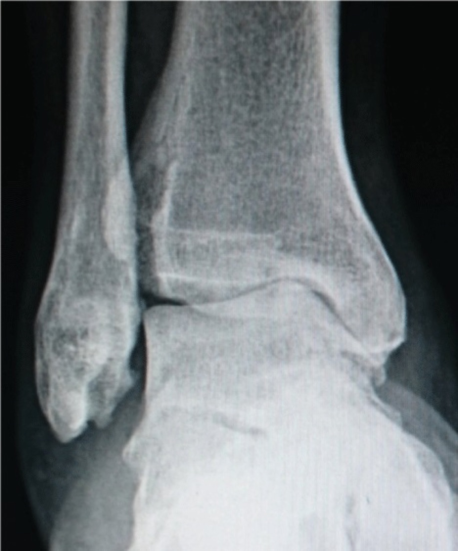
Ankle arthroscopy is a minimally invasive operation in which a camera and fine instruments are introduced into the joint through millimetre-sized incisions (portals). It is most often used for ankle impingement syndrome; removal of loose bodies, treatment of synovitis, cartilage lesions and ankle fusion can also be performed arthroscopically. Because the ankle is a narrow, tight joint, arthroscopy is technically demanding; correct portal placement is critical to protect the superficial nerves around it. With the right indication it offers small incisions, fast recovery and a low infection risk.
Anatomy of the Ankle Joint
The ankle (talocrural joint) is a hinge joint between the tibia, the fibula and the talus. The lower ends of the tibia and fibula form a mortise into which the talus fits.
The joint surfaces are covered with hyaline cartilage. The deltoid ligament stabilises the joint medially, and the anterior talofibular, calcaneofibular and posterior talofibular ligaments stabilise it laterally. Because the ankle carries body weight on a relatively small surface, the load per unit area is higher than in other large joints.
Vessels and nerves run just in front of and behind the joint: in front, the branches of the superficial peroneal nerve and the dorsalis pedis artery; behind, the sural nerve and the tibial nerve. Portals are placed to avoid these structures; injury to these superficial nerves is the most common problem in ankle arthroscopy.
Ankle Impingement Syndrome
Impingement syndrome is pain and restricted movement caused by bony spurs (osteophytes) or thickened soft tissue at the joint margins being pinched during movement. There are two main types according to location.
- Anterior impingement: Osteophytes on the front of the tibia and the talar neck are pinched as the foot is pulled up (dorsiflexion). Common in footballers and dancers ('footballer's ankle'); caused by repetitive microtrauma
- Anterolateral soft-tissue impingement: After repeated sprains, thickened synovial and scar tissue (a meniscoid lesion) forms in the front-outer corner of the joint
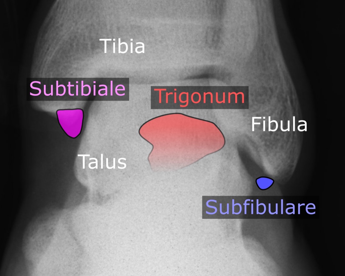
- Posterior impingement: An extra bone behind the talus (os trigonum) or a long posterior process is pinched as the foot is pointed (plantar flexion). Typical in ballet dancers and footballers ('dancer's ankle')
- Previous sprains and repeated overloading are usually at the root of impingement
Other Indications for Ankle Arthroscopy
- Loose bodies: Removal of cartilage or bone fragments
- Osteochondral lesions of the talus: Microfracture, debridement or other cartilage techniques
- Synovial disease: Chronic synovitis, pigmented villonodular synovitis, synovial chondromatosis
- Septic arthritis: Arthroscopic washout and debridement
- Arthroscopy-assisted ligament repair for ankle instability
- Arthrofibrosis: Release of intra-articular adhesions after trauma or surgery
- Assessment of joint-surface step-off after fractures (arthroscopy-assisted fracture surgery)
- Ankle arthrodesis (fusion): Can be performed arthroscopically in advanced arthritis
- Diagnostic arthroscopy: For unexplained chronic ankle pain
Symptoms
- Chronic ankle pain: Worse with activity, partly relieved by rest
- Anterior impingement: Sharp pain at the front when pulling the foot up (squatting, climbing stairs, walking uphill)
- Posterior impingement: Pain at the back when pointing the foot (rising on tiptoe, ballet positions, pressing a pedal)
- Swelling: Particularly after activity
- Restricted movement: Marked limitation of dorsiflexion or plantar flexion
- Catching, locking or clicking: With loose bodies
- Feeling of instability: When there is associated ligament instability
- History of previous sprains: Present in most patients
- Morning stiffness and pain with the first steps after sitting
Diagnosis
The diagnosis is based on a detailed history, examination and imaging. The number of previous sprains, sporting history, which movements provoke the symptoms and previous treatments are reviewed.
Precise localisation of tenderness is important: at the front of the tibia in anterior impingement, the front-outer corner in anterolateral impingement, and beside the Achilles tendon in posterior impingement. Forced dorsiflexion and plantar flexion tests reproduce the pain. Ligament stability tests (anterior drawer, talar tilt) and gait analysis are performed.
Plain radiographs: Standing weight-bearing AP, mortise and lateral views are standard. Anterior osteophytes and the os trigonum are best seen on the lateral view. Special views in forced dorsiflexion may show anterior impingement more clearly.
CT shows the anatomy and extent of osteophytes in detail. MRI is valuable for assessing soft-tissue impingement, synovitis, cartilage lesions, bone oedema and associated ligament pathology. A diagnostic intra-articular local anaesthetic injection helps confirm that the pain arises within the joint.
Non-operative Treatment
Before arthroscopy, at least 3 - 6 months of non-operative treatment is tried where appropriate. Soft-tissue impingement and early cases in particular can benefit significantly.
- Activity modification: Temporarily avoiding movements that provoke impingement
- Physiotherapy: Ankle range of movement, strengthening of the calf and intrinsic foot muscles, proprioception
- Peroneal strengthening: Improves ankle stability and prevents repeated sprains
- Anti-inflammatory medication (NSAIDs): Under medical supervision
- Intra-articular corticosteroid injection: Both diagnostic and therapeutic; ultrasound guidance recommended
- Ankle support or taping: When instability is present
- Heel-raise insoles: May relieve anterior impingement by reducing dorsiflexion
- Footwear adjustment and, if needed, orthotic insoles
Surgical Options
- Arthroscopic debridement: Removal of thickened synovial and scar tissue causing impingement
- Osteophyte resection: Removal of spurs from the front of the tibia and the talar neck
- Os trigonum excision: For posterior impingement — by posterior arthroscopy or an open approach
- Loose body removal
- Synovectomy: For chronic synovitis
- Treatment of cartilage lesions: Microfracture, debridement or appropriate cartilage techniques
- Arthroscopy-assisted ligament repair: When instability is present
- Arthroscopic arthrodesis: Fusion of the joint in advanced arthritis
Expected Outcomes
In well-selected patients, ankle arthroscopy brings a marked reduction in pain and improvement in range of movement. Results are very good, with high patient satisfaction, particularly in isolated impingement with preserved cartilage.
In athletes, return to sport usually takes 2 - 4 months. In contrast, arthroscopy offers limited benefit in patients with marked cartilage damage or advanced arthritis; realistic expectations are important. If instability from repeated sprains is not corrected, impingement may recur.
Surgery & First 3 Weeks
Ankle arthroscopy is performed under spinal/epidural anaesthesia, popliteal nerve block or general anaesthesia. A regional block is effective for pain control in the first 12 - 24 hours. Most cases are day-case surgery.
General Information About the Operation
Radiographs and, if needed, CT and MRI are reviewed; the extent of osteophytes to be removed is planned in advance. Prophylactic antibiotics are given. A tourniquet may be used.
- Duration: Simple debridement 30 - 60 minutes; osteophyte resection and combined procedures 60 - 120 minutes
- Anaesthesia: Spinal/epidural, popliteal block or general anaesthesia
- Hospital stay: Usually day case
- Position: Supine for anterior arthroscopy; prone for posterior arthroscopy
Portals and Nerve Safety
Correct portal placement is the most critical safety step of ankle arthroscopy. The superficial nerves around the joint lie very close to the skin.
- Anteromedial portal: Just medial to the tibialis anterior tendon; the first portal. Close to the saphenous nerve and vein
- Anterolateral portal: Just lateral to the peroneus tertius tendon; branches of the superficial peroneal nerve run right here. Before making the portal, plantar flexing and inverting the foot can make the nerve visible through the skin so it can be marked
- Posterolateral portal: Just lateral to the Achilles tendon; close to the sural nerve
- Posteromedial portal: In posterior arthroscopy, just medial to the Achilles tendon; the portal requiring most care because of the tibial nerve and vessels
- Only the skin is incised; the deeper tissues are spread bluntly with a small clamp — a technique that markedly reduces nerve injury
Opening the Joint Space
- Non-invasive traction: A soft strap on the foot applies controlled distraction to widen the joint; needed to reach the back of the joint and cartilage lesions
- Dorsiflexion technique: In anterior impingement surgery, work is done without traction with the foot pulled up; this relaxes the anterior capsule and creates working space
- Invasive (pin) traction is rarely used today; its complication risk is higher
- Traction time is kept as short as possible
Anterior Arthroscopy — Surgical Technique
- A 2.7 mm or 4.0 mm camera is introduced through the anteromedial portal and the joint is inspected systematically
- Instruments are introduced through the anterolateral portal
- Synovitis and thickened soft tissue (meniscoid lesion) are removed with a shaver
- Osteophytes on the front of the tibia and the talar neck are removed with a burr; the extent of resection can be confirmed with fluoroscopy
- After resection the ankle is dorsiflexed dynamically to confirm on the table that impingement has been relieved
- Loose bodies are removed with a grasper
- Any cartilage lesion is assessed and microfracture performed if needed
- Under-resection leaves symptoms; over-resection may compromise joint stability
Posterior Arthroscopy and Os Trigonum Excision
- The patient lies prone with the foot over the end of the table
- Posterolateral and posteromedial portals are made on either side of the Achilles tendon
- Work is kept lateral to the flexor hallucis longus (FHL) tendon — this tendon is the anatomical boundary protecting the tibial nerve and vessels, and the most important safety rule of posterior arthroscopy
- The posterior capsule and fat are cleared to gain a view
- The os trigonum or a long posterior process of the talus is freed and removed
- The FHL tendon sheath is released if needed (a common associated problem in dancers)
- Relief of impingement in plantar flexion is tested on the table
Discharge and Weight-bearing
Patients usually go home the same day. The weight-bearing protocol depends on the procedure: early weight-bearing is possible after simple debridement and osteophyte resection, whereas a period of non-weight-bearing is needed after cartilage procedures.
- Simple debridement / synovectomy: Weight-bearing as tolerated, usually in a normal shoe
- Osteophyte resection (anterior impingement): Weight-bearing as tolerated; early movement encouraged
- Os trigonum excision: Weight-bearing as tolerated; plantar flexion exercises start early
- After microfracture: 4 - 6 weeks non-weight-bearing or partial weight-bearing
- With added ligament repair: Protection in a boot and partial weight-bearing according to protocol
- Early movement: Important after this operation to prevent intra-articular adhesions
- Ice 3 - 4 times a day and elevation above heart level. Analgesics, anti-inflammatory medication, stomach protection and, if non-weight-bearing, anticoagulation are prescribed. Stitches are removed at 10 - 14 days
Early Postoperative Period (First 3 Weeks)
The aims in this period are to control swelling, regain range of movement early, heal the portal wounds and monitor the superficial nerves. Swelling around the ankle is stubborn and directly affects recovery.
- Swelling control: Keeping the leg above heart level is particularly important after this operation; full range cannot be regained until swelling subsides
- Ice: 15 - 20 minutes 3 - 4 times a day
- Early range-of-movement exercises: Active and active-assisted dorsiflexion and plantar flexion; prevent adhesions
- Weight-bearing: According to protocol; early full weight-bearing is encouraged after simple procedures
- Wound care: Portal sites kept dry for 10 - 14 days; discharge, redness and warmth monitored. Ankle portals are prone to leaking joint fluid
- Nerve monitoring: Numbness on the top of the foot (superficial peroneal), the outer edge (sural) or the inner edge (saphenous) should be reported
- Compression stockings
- Thromboprophylaxis: Anticoagulation and toe exercises during non-weight-bearing
Follow-up
- Days 10 - 14: Removal of stitches and wound check
- Week 6: Range-of-movement and weight-bearing assessment
- Month 3: Strength and function assessment, return-to-sport decision
- Month 6: Outcome assessment
Risks & Complications
Ankle arthroscopy is a safe operation when performed with correct technique, and serious complications are rare. Its most characteristic risk is injury to the superficial nerves around the joint when portals are made, because these nerves lie very close to the skin.
Risks Specific to Non-operative Treatment
- Persistent symptoms and continued activity restriction
- Progression of impingement: Enlarging osteophytes and increasing restriction of movement
- Progression of cartilage damage and development of ankle arthritis in the long term
- Adverse effects of repeated corticosteroid injections on cartilage and tendon
- Untreated instability leading to further sprains and additional damage
Surgical Risks — Common
- Superficial nerve injury: The most common complication of this operation. The superficial peroneal nerve (anterolateral portal), sural nerve (posterolateral portal) and saphenous nerve (anteromedial portal) may be affected, causing numbness or tingling on the top or edges of the foot; usually temporary
- Temporary tenderness and numbness at the portal sites
- Superficial portal infection (usually treated with antibiotics)
- Prolonged swelling: May last 6 - 12 months in this region
- Postoperative pain and swelling
- Temporary ankle stiffness — improves with physiotherapy
- Leakage of joint fluid from a portal: Usually stops on its own
Surgical Risks — Less Common
- Permanent neuropathy or painful neuroma: A painful nodule at the end of an injured superficial nerve; may be difficult to treat
- Deep joint infection (under 1%): Requires arthroscopic washout and antibiotics
- Deep vein thrombosis (DVT) and pulmonary embolism: Increased risk during non-weight-bearing
- Tibial nerve or vessel injury: In posterior arthroscopy if work strays medial to the FHL tendon; very rare but serious
- Cartilage injury: Scuffing of the joint surface when instruments are introduced
- Inadequate osteophyte resection: Persistent symptoms
- Over-resection: Compromised joint stability
- Arthrofibrosis (intra-articular adhesions): Restricted movement
- Complex regional pain syndrome (CRPS): Rare, but reported more often after ankle surgery than in other regions
- Instrument breakage with retained fragments (very rare)
Risk Factors
- Advanced ankle arthritis: The expected benefit of arthroscopy falls markedly
- Widespread cartilage damage
- Uncorrected ligament instability: Leads to recurrent impingement
- Malalignment (varus or valgus ankle)
- Smoking and poorly controlled diabetes
- Peripheral vascular disease and venous insufficiency (worsen swelling)
- Obesity
- Previous ankle surgery and scar tissue
- Poor adherence to rehabilitation
Contact Your Doctor Immediately If
- Pain or swelling suddenly increases
- You have a temperature above 38 °C
- There is discharge, a bad smell or an opening at a portal site
- Numbness, tingling or weakness in the foot or toes is progressing
- Your toes become blue, cold or pale
- There is firm, one-sided swelling and redness of the calf (suspected DVT)
- You develop shortness of breath or chest pain (suspected pulmonary embolism)
- You have burning, disproportionate pain and extreme sensitivity (suspected CRPS)
Long-term Outcomes
In patients with preserved cartilage and isolated impingement, the results of arthroscopy are very good; most become pain-free and return to sport. Results of anterior impingement surgery are generally somewhat more predictable than those for posterior impingement.
Osteophytes can re-form over time, particularly in athletes who continue the same loading pattern. In patients with advanced cartilage damage, arthroscopy may give temporary relief but does not halt the progression of arthritis; fusion or, in selected patients, ankle replacement is then considered. Correcting associated instability is an important part of long-term success.
Rehabilitation
Rehabilitation after ankle arthroscopy has two priorities: controlling swelling and regaining range of movement early. Swelling in this region is stubborn and full range cannot be regained until it resolves, so elevation and ice matter as much as exercise. The programme differs markedly according to the procedure performed.
After Simple Debridement and Osteophyte Resection
- Week 0 - 1: Weight-bearing as tolerated; crutches briefly if needed. Elevation, ice and compression stockings. Active ankle exercises (dorsiflexion-plantar flexion) several times a day.
- Weeks 1 - 3: Walking distance increased gradually. Range-of-movement work intensifies. Isometric exercises for the calf and intrinsic foot muscles. Scar massage once healed.
- Weeks 3 - 6: Resistance-band strengthening (dorsiflexion, plantar flexion, inversion, eversion). Balance and proprioception exercises. Stationary cycling.
- Weeks 6 - 12: Strengthening increased; single-leg balance work. Swimming. Light running on flat ground (if criteria are met).
- Months 3 - 4: Agility and sport-specific work; gradual return to sport.
After Os Trigonum Excision (Posterior Impingement)
- Week 0 - 1: Weight-bearing as tolerated. Elevation and ice. Early plantar flexion exercises are important (to prevent adhesions).
- Weeks 1 - 4: Range of movement, particularly plantar flexion, increased gradually. If the FHL tendon was released, big-toe movement exercises are added.
- Weeks 4 - 8: Strengthening and proprioception. Calf strengthening.
- Months 2 - 4: Gradual return to pointe work in dancers and to kicking in footballers.
- Return to sport in this group usually takes 2 - 4 months.
When a Microfracture or Cartilage Procedure Is Added
- Weeks 0 - 4/6: Non-weight-bearing or partial weight-bearing; crutches. Early passive and active-assisted movement supports cartilage healing.
- Weeks 4 - 8: Gradual weight-bearing. Active movement and light strengthening.
- Months 2 - 4: Full weight-bearing; strengthening and balance work intensify.
- Months 4 - 6: Light running begins.
- Months 6 - 12: Gradual return to sport; cartilage maturation takes time.
Swelling Control — Particularly Important After This Operation
- Keep the leg above heart level: For most of the day in the first 2 weeks, and as much as possible thereafter
- Ice: 15 - 20 minutes 3 - 4 times a day; consistently in the first 2 weeks
- Compression stockings: Regular use for 3 - 6 months is recommended
- Ankle pumping exercises: Reduce swelling and the risk of clots
- Avoid standing for long periods and letting the leg hang
- Swelling may persist for 6 - 12 months in this region; this is expected, but increasing swelling should be reported
Return-to-sport and Activity Criteria
- Full, pain-free ankle range of movement
- Calf strength at least 85 - 90% of the other leg
- Adequate repetitions on the single-leg heel raise test
- Symmetrical single-leg balance (30 seconds with eyes closed)
- 90% symmetry on single-leg hop tests
- Pain-free running and changes of direction
- No swelling or pain after activity
- Pain-free completion of sport-specific movements
- Approval from the surgeon and physiotherapist
Recommended and Avoided Activities
- Recommended: Stationary cycling, swimming (once healed), elliptical trainer, walking on flat ground
- Recommended: Resistance-band strengthening, balance board, proprioception work
- Avoid in the first 6 weeks: Jumping, running, sudden changes of direction
- Avoid in the first 3 months: Walking and running on uneven ground, prolonged standing
- After a cartilage procedure: High-impact activity restricted for 6 - 12 months
- Avoid: Long walks in unsupportive shoes; surfaces with a high risk of spraining
Lifestyle and Recovery Advice
- Keep the leg above heart level as much as possible in the first weeks — the simplest measure that most speeds recovery after this operation
- Do your home exercise programme every day; early range-of-movement work prevents adhesions
- Wear compression stockings regularly (3 - 6 months)
- Do not smoke; it impairs tissue healing
- If you have diabetes, keep your blood glucose tightly controlled
- Healthy weight: The ankle carries some of the highest loads per unit area of any joint
- If you have ankle instability, continue peroneal strengthening for life; repeated sprains are the most common cause of impingement
- If you are an athlete, review your movement technique and loading pattern with your physiotherapist
- Use appropriate footwear and, if needed, insoles
- Attend regular follow-up (week 2, week 6, month 3, month 6)
What to Expect
After simple debridement and osteophyte resection, return to daily activities usually takes 2 - 4 weeks. Return to sport takes 2 - 4 months. When a cartilage procedure is added, these times are considerably longer (return to sport 6 - 12 months).
Mild swelling, tenderness at the portal sites and stiffness after activity may persist for 6 - 12 months; this is expected and settles with time. A small patch of numbness on the top or edge of the foot may be permanent. The care given to swelling control directly determines the speed of recovery after this operation.
Is This Treatment Right for You?
Every patient's condition is different. After a detailed examination and imaging, the treatment plan that suits you best will be prepared.
Would you like more information about Ankle Arthroscopy?
Book Appointment