Hip Arthroscopy
Arthroscopic treatment of femoroacetabular impingement and labral tears
Overview
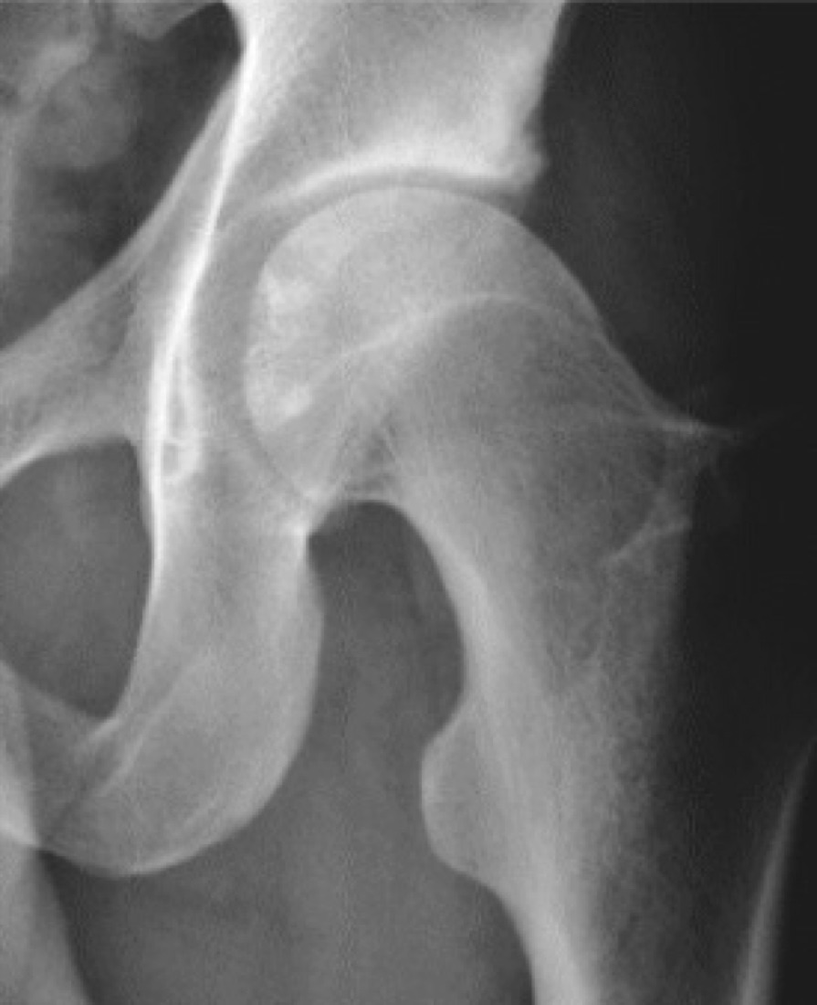
Hip arthroscopy is a minimally invasive operation in which a camera and specialised instruments are introduced into the hip joint through small incisions. It is most often used for femoroacetabular impingement (FAI) and the labral tears that accompany it. The hip is one of the deepest joints in the body and is surrounded by powerful muscles; hip arthroscopy is therefore technically more demanding than knee or shoulder arthroscopy, and controlled distraction of the leg (traction) is needed to gain access to the joint. Correct patient selection is the single most important determinant of outcome: hip arthroscopy does not deliver the expected benefit in patients with advanced osteoarthritis.
Anatomy of the Hip and Labrum
The hip is a ball-and-socket joint between the spherical femoral head and the cup-shaped acetabulum of the pelvis. The joint surfaces are covered with hyaline cartilage.
The labrum is a ring of fibrocartilage, triangular in cross-section, around the rim of the acetabulum. It deepens the socket to grip the femoral head, keeps joint fluid inside to create a suction-seal effect, and spreads load over a wider area. When the labrum tears, this seal is lost; pressure distribution within the joint changes and load on the cartilage rises.
The joint is also enclosed by a strong capsule and the iliofemoral ligament (ligament of Bigelow). The capsule plays an important role in hip stability; appropriate repair of the capsule opened during arthroscopy is important to prevent postoperative instability.
Femoroacetabular Impingement (FAI)
FAI is abnormal contact between the femoral head-neck junction and the acetabular rim during movement. This repeated collision gradually damages the labrum and adjacent cartilage. There are three types:
- Cam type: A bony bump at the femoral head-neck junction disrupts the sphericity of the head. When the knee is brought to the chest and turned inwards, the bump slides under the labrum and strips the cartilage. More common in young men and athletes
- Pincer type: Excessive acetabular coverage of the femoral head; the labrum is pinched between the femoral neck and the acetabular rim. More common in middle-aged women
- Mixed type: Both mechanisms together; the most common type
- Relationship to sport: More common in football, hockey, martial arts, ballet and gymnastics, which repeatedly force the hip into deep flexion and rotation
Other Indications for Hip Arthroscopy
- Isolated labral tears (traumatic or degenerative)
- Loose bodies: Cartilage or bone fragments
- Ligamentum teres injuries
- Cartilage lesions: Microfracture or other cartilage repair techniques
- Synovial disease: Pigmented villonodular synovitis (PVNS), synovial chondromatosis
- Septic arthritis: Arthroscopic washout and debridement
- Psoas tendon impingement (internal snapping hip) and tendon release when needed
- Gluteus medius/minimus tendon pathology (peritrochanteric space surgery)
- Iliotibial band release for external snapping hip
Symptoms
- Groin pain: The most typical complaint; patients often cup the hip with thumb and index finger in a 'C' shape when describing it (the 'C sign')
- Deep, dull pain: At the front of the hip and in the groin, sometimes radiating to the front of the thigh and the side of the hip
- Worse with activity: Prolonged sitting, driving, tying shoelaces, getting in and out of a car, squatting and climbing stairs
- Catching, clicking or locking: With a labral tear or loose body
- Restricted movement: Particularly hip flexion and internal rotation
- Reduced performance in athletes: Difficulty with sudden acceleration, changes of direction and kicking
- Night pain and stiffness on standing after prolonged sitting
- Usually insidious onset: Most patients have no clear history of injury
Diagnosis
The diagnosis is based on a detailed history, careful examination and imaging considered together. Differential diagnosis matters, because hip pain can be confused with lumbar disc disease, sacroiliac joint problems, inguinal hernia and adductor tendinopathy.
The most valuable examination finding is the FADIR (Flexion-Adduction-Internal Rotation) test: sharp groin pain when the hip is flexed and rotated inwards suggests impingement. The FABER test, measurement of hip range of movement (particularly loss of internal rotation), gait analysis and assessment of neighbouring regions are also performed.
Plain radiographs: A standing AP pelvis, a lateral view (Dunn or cross-table lateral) and, if needed, a false-profile view. The alpha angle is measured for cam lesions, the crossover sign and lateral centre-edge angle (LCEA) for pincer lesions, and the joint space is assessed — the most critical part of the surgical decision.
MR arthrography (MRI with contrast injected into the joint) is superior to standard MRI for showing labral tears. CT with 3D reconstruction shows bone morphology and the extent of bone to be resected in detail and is used for planning. A diagnostic intra-articular local anaesthetic injection is valuable in confirming whether the pain truly arises from within the joint.
Non-operative Treatment
Before hip arthroscopy, at least 3 - 6 months of non-operative treatment is tried in suitable patients. In some patients symptoms are adequately controlled this way.
- Activity modification: Avoiding deep squatting, prolonged sitting and movements that provoke impingement
- Physiotherapy: Strengthening of the hip and core muscles, balancing of the muscles around the hip, movement-pattern retraining
- Strengthening of the gluteal muscles and deep hip rotators: Improves the movement of the femoral head within the socket
- Anti-inflammatory medication (NSAIDs): Under medical supervision
- Intra-articular corticosteroid injection: Both therapeutic and diagnostic; given under ultrasound or fluoroscopic guidance
- Weight control
- Technique adjustment in athletes: Review of loading patterns and movement technique
- Response to non-operative treatment may be limited, particularly when bone morphology is markedly abnormal
Surgical Options
- Labral repair: Reattachment of the torn labrum to the acetabular rim with suture anchors; today, repairing the labrum is preferred over removing it because preserving the seal matters in the long term
- Labral reconstruction: Reconstruction with a graft when the labrum is too damaged to repair
- Femoral osteoplasty (cam resection): Shaving the bony bump at the head-neck junction to restore sphericity
- Acetabular rim trimming (pincer resection): Controlled removal of the over-covering acetabular rim
- Cartilage treatment: Debridement, microfracture or other cartilage techniques according to the lesion
- Capsular repair (capsuloplasty): Closure of the opened capsule; important to prevent instability
- Loose body removal and synovectomy
- Psoas or iliotibial band release: For snapping hip syndrome
Who Is — and Who Is Not — a Candidate
In this operation, patient selection is more decisive than surgical technique. In a well-selected patient the results are very good; in a poorly selected patient they are disappointing.
- Suitable: Young and middle-aged patients with a preserved joint space and no significant osteoarthritis
- Suitable: Patients who have not responded to non-operative treatment and have FAI morphology and a labral tear on imaging
- Suitable: Patients who respond clearly to a diagnostic intra-articular injection (confirming the pain arises within the joint)
- Not suitable: Advanced hip osteoarthritis (joint space below 2 mm) — arthroscopy does not help; hip replacement should be considered
- Not suitable: Advanced age with significant cartilage loss
- Requires careful assessment: Hip dysplasia (a shallow socket) — arthroscopy alone may worsen instability; a periacetabular osteotomy may be needed
- Requires careful assessment: Generalised ligamentous laxity
Expected Outcomes
In well-selected patients, hip arthroscopy brings a marked reduction in pain and improvement in function. Most patients return to daily activities within 3 - 6 months; return to sport takes 4 - 9 months in athletes.
Recovery is clearly slower after hip arthroscopy than after knee arthroscopy; knowing this from the outset matters for realistic expectations. Long-term results are good in young patients with well-preserved cartilage. In patients with advanced cartilage damage, symptoms may improve partially, but hip replacement may be needed over time.
Surgery & First 3 Weeks
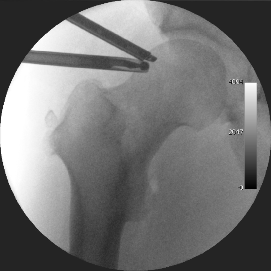
Hip arthroscopy is performed under spinal/epidural or general anaesthesia on a special traction table, with fluoroscopic guidance. Controlled traction of the leg to gain access to the joint is the most distinctive technical feature of this operation.
General Information About the Operation
Plain radiographs, MR arthrography and, if needed, 3D CT are reviewed; the limits of the bone to be resected are planned in advance. Prophylactic antibiotics are given and thromboprophylaxis is planned.
- Duration: 90 - 150 minutes (depending on the procedures performed)
- Anaesthesia: Spinal/epidural or general; full muscle relaxation is needed for traction
- Hospital stay: Usually day case or one night
- Traction time: Kept as short as possible (usually under 2 hours); prolonged traction increases the risk of nerve injury
Positioning and Traction
- The patient is placed supine or lateral on the traction table
- A wide, well-padded perineal post is used, chosen as wide as possible to reduce pressure on the pudendal nerve
- Controlled traction is applied to the operated leg to create an 8 - 10 mm gap between the femoral head and acetabulum, confirmed by fluoroscopy
- Traction is applied only while working in the central compartment; it is released for the peripheral compartment (femoral neck region)
- Keeping traction time short is the most important safety rule of this operation
Portals and Joint Access
- Anterolateral portal: The first and safest portal, entered under fluoroscopy at the anterosuperior border of the greater trochanter
- Anterior or modified mid-anterior portal: Used for instruments; requires care because of the nearby lateral femoral cutaneous nerve
- Posterolateral portal: Added when needed
- Needle entry, guidewire placement and cannula advancement are all done under fluoroscopic control
- Interportal capsulotomy: The capsule is opened in a controlled way to enlarge the working space and repaired at the end
Central Compartment — Labral and Cartilage Surgery
- Under traction the joint is inspected: labrum, acetabular cartilage, femoral head cartilage and ligamentum teres
- The type and location of the labral tear are defined; areas of cartilage delamination are assessed
- If a pincer lesion is present, the acetabular rim is trimmed in a controlled way; this may require temporary detachment of the labrum
- Labral repair: The labrum is reattached with suture anchors placed on the acetabular rim; sutures are passed to preserve the seal
- If the labrum cannot be repaired, it is reconstructed with a graft
- Cartilage lesions: Debridement, microfracture or an appropriate cartilage technique
- Loose bodies are removed; partial synovectomy is added if there is synovitis
Peripheral Compartment — Femoral Osteoplasty
- Traction is released and the hip is flexed to expose the femoral head-neck junction
- The cam lesion is removed in a controlled way with a burr; the aim is to restore the spherical shape of the head
- The limits of resection are confirmed with fluoroscopy and dynamic movement of the hip; relief of impingement is tested on the table
- Under-resection leaves symptoms; over-resection risks femoral neck fracture — getting this balance right is the most critical step
- Preservation of the retinacular vessels: The femoral head's blood supply depends on them, and the resection limit is set accordingly
Capsular Repair and Closure
- The opened capsule is sutured; this is particularly important to prevent instability in patients with laxity or borderline dysplasia
- Portals are closed; local anaesthetic may be injected into the joint
- Traction is released and a neurovascular examination is performed
Discharge and Weight-bearing
Patients usually go home the same day or the next day. The weight-bearing and movement protocol after surgery depends on the procedures performed, and adherence directly affects the result.
- Isolated labral repair and osteoplasty: Usually 2 - 4 weeks of partial weight-bearing (foot-flat with crutches)
- After microfracture: 6 - 8 weeks of partial weight-bearing
- After capsular repair: Excessive external rotation and extension are limited for the first 3 weeks
- Movement restrictions: Hip flexion is usually limited to 90° for the first 2 - 3 weeks; active hip flexion is limited early (to prevent psoas tendinitis)
- Some protocols use a hip brace or anti-rotation splint
- Continuous passive motion (CPM) or a stationary bike (no resistance) is recommended early to prevent adhesions
- Heterotopic ossification prophylaxis: Some centres give anti-inflammatory medication for 2 - 4 weeks
- Ice 3 - 4 times a day; analgesics, anti-inflammatory medication, stomach protection and anticoagulation are prescribed. Stitches are removed at 10 - 14 days
Early Postoperative Period (First 3 Weeks)
The aims in this period are to protect the repaired labrum and capsule, prevent intra-articular adhesions, control swelling and slow muscle wasting. The most common mistake in the early phase after hip arthroscopy is starting active hip flexion exercises too soon and irritating the psoas tendon.
- Adherence to the weight-bearing protocol: The period of partial weight-bearing is critical, particularly after microfracture
- Early passive movement: CPM or a stationary bike (no resistance, short sessions) is the most effective way to prevent adhesions
- Passive rotation exercises: Gentle inward and outward rotation of the hip while lying on the back
- Active hip flexion is limited: Straight-leg raises and active knee-to-chest exercises are avoided for the first 3 weeks (to prevent psoas irritation)
- Isometric exercises: Gluteal, quadriceps and transversus abdominis contractions
- Swelling control: Ice, elevation and compression stockings
- Wound care: Portal sites kept dry for 10 - 14 days; discharge, redness and warmth monitored
- Thromboprophylaxis: Anticoagulation, compression stockings and ankle pumps
- Neurological monitoring: Numbness in the perineum, loss of sensation on the top of the foot or tingling on the outer thigh should be reported
Follow-up
- Days 10 - 14: Removal of stitches and review of early rehabilitation
- Week 6: Weight-bearing decision and range-of-movement check
- Month 3: Strength assessment and activity progression
- Month 6 and year 1: Return-to-sport assessment and follow-up imaging if needed
Risks & Complications
Hip arthroscopy is a minimally invasive operation and the rate of serious complications is low. However, it carries specific risks not seen in other joint arthroscopies, most of which arise from the traction applied to the leg.
Traction-related Risks (Specific to This Operation)
- Pudendal nerve injury: Caused by pressure from the perineal post; may cause perineal numbness and sexual dysfunction. Usually temporary but occasionally permanent. The risk is reduced with a wide, well-padded post
- Sciatic and femoral nerve traction neurapraxia: Numbness and temporary weakness in the leg; the risk rises with longer traction time
- Pressure injury to the perineal skin and swelling of the scrotum/labia
- Pressure injury around the ankle and foot from the traction boot
- All these risks are markedly reduced by keeping traction time short
Common Risks (Under 5%)
- Lateral femoral cutaneous nerve injury: Numbness or tingling on the outer thigh; the most common nerve problem because of the anterior portal. Usually temporary
- Superficial portal infection (usually treated with antibiotics)
- Postoperative pain, swelling and bruising
- Fluid extravasation: Arthroscopic fluid leaking into surrounding tissues; usually resolves spontaneously
- Temporary hip stiffness
- Psoas tendinitis: Linked to early active hip flexion exercises; causes pain at the front of the groin
- Prolonged swelling and a feeling of fullness in the groin (may last 6 - 12 months)
Less Common Risks
- Heterotopic ossification: Bone formation in soft tissue; may restrict movement. The risk is reduced with anti-inflammatory prophylaxis
- Femoral neck fracture: After excessive femoral osteoplasty; the risk rises with early weight-bearing. The resection limit and weight-bearing protocol are therefore set with great care
- Avascular necrosis of the femoral head: Due to damage to the retinacular vessels; very rare but serious
- Joint instability: After failure to repair the capsule or excessive rim resection; the risk is high in borderline dysplasia
- Deep joint infection (under 1%)
- Deep vein thrombosis (DVT) and pulmonary embolism
- Intra-abdominal fluid extravasation: Very rare but serious; presents with abdominal distension and low blood pressure
- Instrument breakage with retained fragments (very rare)
- Chondrolysis (rapid cartilage loss): Very rare
Causes of Surgical Failure
The most common reasons for persistent symptoms after hip arthroscopy relate more to patient selection than to technical problems.
- Poor patient selection: Arthroscopy in a patient with advanced osteoarthritis does not deliver the expected benefit — the most common cause of failure
- Inadequate bone resection: Incomplete correction of the cam or pincer lesion; impingement persists
- Over-resection: Risk of instability or femoral neck fracture
- Missed dysplasia: Arthroscopy alone in a patient with a shallow socket may make things worse
- Failure to repair the capsule: Leads to instability, particularly in patients with laxity
- Missed associated lumbar, sacroiliac or groin pathology
- Poor adherence to rehabilitation
Risk Factors
- Advanced age and existing cartilage damage
- Joint-space narrowing (below 2 mm) — a relative contraindication
- Hip dysplasia and generalised ligamentous laxity
- Long traction time
- Obesity (both surgical difficulty and outcome)
- Smoking and poorly controlled diabetes
- Previous hip surgery
- High expectations with unrealistic goals
Contact Your Doctor Immediately If
- Groin or hip pain suddenly increases, especially sharp pain on weight-bearing (suspected femoral neck fracture)
- You have a temperature above 38 °C
- There is discharge, a bad smell or an opening in the wound
- You have perineal numbness or a change in urinary or sexual function (suspected pudendal nerve injury)
- Numbness, tingling or weakness in the leg or foot is progressing
- There is firm, one-sided swelling and redness of the leg (suspected DVT)
- You develop shortness of breath or chest pain (suspected pulmonary embolism)
- You have abdominal tightness, distension and weakness (suspected fluid extravasation — urgent)
Long-term Outcomes
In well-selected patients, hip arthroscopy provides a marked reduction in pain and a lasting improvement in function, with high patient satisfaction. Repairing rather than removing the labrum has clearly improved long-term results.
In young patients with well-preserved cartilage the results last for many years. In contrast, when advanced cartilage damage is found at surgery, symptoms may improve partially, but hip replacement may be needed over time. Research continues into how far correcting FAI prevents hip osteoarthritis in the long term; the most honest information for the patient is that the operation aims to relieve current symptoms.
Rehabilitation
Rehabilitation after hip arthroscopy is clearly longer and more gradual than after knee arthroscopy. The joint is deep, the muscles are powerful and the repaired labrum and capsule need time to heal. The programme has two special rules: passive movement early on to prevent intra-articular adhesions, and avoiding active hip flexion so as not to irritate the psoas tendon.
Phase 1 — Protection and Movement (0 - 4 Weeks)
- Weight-bearing: Partial weight-bearing with crutches according to the surgeon's protocol (2 - 4 weeks after labral repair, 6 - 8 weeks after microfracture)
- Passive movement: CPM or a stationary bike (no resistance, 20 minutes once or twice a day) — the most effective way to prevent adhesions
- Passive rotation exercises: Gentle internal and external rotation
- NO active hip flexion: Straight-leg raises and active knee-to-chest are avoided for the first 3 weeks because they irritate the psoas
- Isometric work: Gluteal, quadriceps and core contractions
- Movement limits: Hip flexion usually limited to 90°; external rotation and extension limited according to the capsular repair
- Ice, elevation and swelling control
Phase 2 — Range of Movement and Activation (4 - 8 Weeks)
- Gradual progression to full weight-bearing; weaning off crutches and correcting gait
- Movement restrictions lifted; full range is the goal
- Gradual introduction of active hip movement (once psoas irritation has been excluded)
- Gluteus medius and maximus activation: Bridges, side-lying leg raises, clamshells
- Core stabilisation work
- Pool walking and water exercise (once the wound has healed)
- Stationary bike resistance increased gradually
Phase 3 — Strengthening (2 - 4 Months)
- Closed-chain strengthening: Mini squats, leg press, step-ups
- Resisted gluteal work; single-leg control exercises
- Balance and proprioception: Single-leg balance, balance board
- Targeted strengthening of hip abductors and external rotators
- Elliptical trainer and walking distance increased gradually
- Swimming (once the wound has fully healed)
Phase 4 — Functional Training and Return to Sport (4 - 9 Months)
- Months 4 - 5: Light running on flat ground (if criteria are met)
- Months 5 - 6: Gradual addition of agility, change-of-direction and plyometric exercises
- Months 6 - 9: Sport-specific drills and return to full training
- In contact and high-rotation sports (football, hockey, martial arts), return may take until month 9
- The athlete's movement patterns and loading technique are reviewed and adjusted to prevent recurrence
Return-to-sport and Activity Criteria
- Pain-free hip range of movement close to the other side
- Hip abductor and extensor strength at least 85 - 90% of the other side
- No inward collapse of the knee during single-leg squat and landing
- 90% symmetry on single-leg hop tests
- Pain-free running and changes of direction
- No groin pain or swelling after activity
- Pain-free completion of sport-specific movements
- Approval from the surgeon and physiotherapist
Recommended and Avoided Activities
- Recommended: Stationary cycling, swimming, pool walking, elliptical trainer, walking on flat ground
- Recommended: Gluteal and core strengthening, controlled Pilates
- Avoid in the first 3 weeks: Active hip flexion (straight-leg raises), prolonged sitting
- Avoid in the first 3 months: Deep squatting, sitting cross-legged, stretches that force the hip
- Avoid in the first 6 months: Sudden changes of direction, pivoting, jumping
- Avoid: Heavy deep squats and yoga poses that force the hip to extreme angles
Lifestyle and Recovery Advice
- Be patient: Recovery after hip arthroscopy is measured in months; slower-than-expected progress in the first 3 months is normal
- Do your home exercise programme every day; gluteal work in particular directly determines the result
- Avoid active hip flexion exercises in the early phase; if pain starts at the front of the groin, tell your physiotherapist
- Avoid prolonged sitting; get up and move every hour — sitting mimics the impingement position
- Healthy weight: Directly reduces load on the hip
- Do not smoke; it impairs tissue and bone healing
- If you have diabetes, keep your blood glucose tightly controlled
- Follow the weight-bearing programme strictly; excessive early load after osteoplasty carries a risk of femoral neck fracture
- If you are an athlete, review your movement technique with your physiotherapist; if the same loading pattern continues, the problem may recur
- Attend regular follow-up (week 6, month 3, month 6, year 1)
What to Expect
Return to daily activities usually takes 6 - 12 weeks. Running resumes at 4 - 5 months and full return to sport at 6 - 9 months. Full functional recovery may take 9 - 12 months.
A mild feeling of fullness in the groin, stiffness after prolonged sitting and mild pain after activity may persist for 6 - 12 months; this is expected and settles with time. A small patch of numbness on the outer thigh may be permanent. Knowing from the outset that recovery is clearly slower than after knee arthroscopy prevents unnecessary worry.
Is This Treatment Right for You?
Every patient's condition is different. After a detailed examination and imaging, the treatment plan that suits you best will be prepared.
Would you like more information about Hip Arthroscopy?
Book Appointment