Achilles Tendon Rupture
Surgical and non-operative treatment of Achilles tendon rupture with a return-to-sport programme
Overview
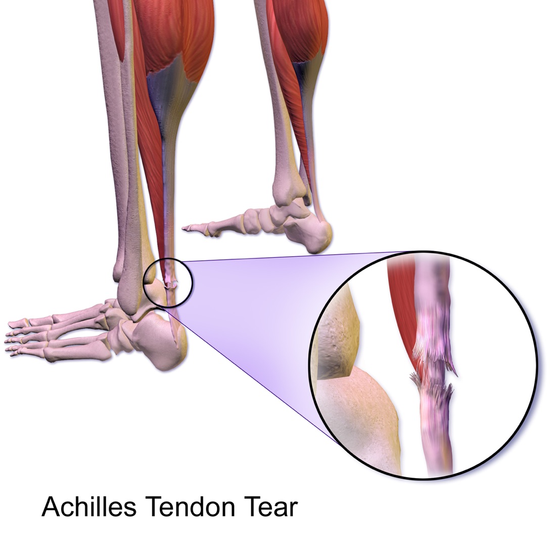
The Achilles tendon connects the calf muscles (gastrocnemius and soleus) to the heel bone and is the thickest and strongest tendon in the body. It carries several times body weight during walking, running and jumping. Achilles tendon rupture occurs most often in men aged 30 - 50 who do not exercise regularly but occasionally take part in high-intensity activity. The tear typically occurs 2 - 6 cm above the insertion on the heel, the region with the poorest blood supply. Treatment may be surgical or non-operative; thanks to modern functional rehabilitation protocols, the difference in outcome between the two has narrowed considerably.
Anatomy of the Achilles Tendon
The Achilles tendon is the common tendon formed by the gastrocnemius and soleus muscles at the back of the calf and inserts on the back of the heel bone (calcaneus). It produces plantar flexion — pointing the foot downwards; lifting the heel off the ground during walking depends on it.
Blood supply varies along the tendon. The region 2 - 6 cm above the insertion is the most poorly vascularised (the watershed zone), and the great majority of ruptures occur here. The same poor blood supply is why healing is slow.
The thin paratenon sheath around the tendon contributes to its nutrition and gliding; preserving it during surgery is important for healing.
Mechanisms of Rupture
- Sudden acceleration or change of direction: Push-off or jumping in tennis, basketball, football or badminton
- Unprepared activity: A person who does not exercise regularly suddenly taking part in intense activity (the 'weekend athlete')
- Forced dorsiflexion of the ankle: Missing a step on stairs or a kerb; stepping into a hole
- Direct blow: A blow to the tensed tendon from behind (less common)
- Degenerative background: Most ruptures occur in a tendon that already has degeneration (tendinopathy), often without prior symptoms
- Medication: Fluoroquinolone antibiotics and corticosteroids increase the risk of tendon rupture
Symptoms
- Sudden, sharp pain: Most patients describe feeling as if they had been 'kicked' or 'hit by a stone' at the back of the heel
- Audible snap: A 'pop' may be heard at the moment of rupture
- Inability to rise on tiptoe: A single-leg heel raise cannot be performed on the injured side — the most valuable clinical sign
- Abnormal gait: Walking is possible but push-off power is lost; the patient limps
- Palpable gap: A depression felt over the tendon (more obvious in the first hours; less so as swelling increases)
- Swelling and bruising: Behind the heel and in the lower calf
- Misleadingly reassuring sign: The toes can still be pointed downwards because other muscles do this, so the rupture can be missed
Diagnosis
The diagnosis is largely clinical. The circumstances of the injury, whether a snap was heard, sporting history and any use of fluoroquinolones or corticosteroids are reviewed.
The tendon is palpated along its length for a gap. The Thompson (Simmonds) test is the most valuable sign: with the patient lying face down, squeezing the calf normally makes the foot point downwards; in a complete rupture this does not happen. Inability to rise on tiptoe on one leg supports the diagnosis.
Ultrasound shows whether the rupture is complete or partial, where it is and how far apart the tendon ends are; its ability to examine the tendon dynamically is an advantage. MRI gives detailed information in partial ruptures, chronic cases and surgical planning. Plain radiographs do not show a tendon rupture; they are taken to exclude an associated calcaneal fracture or bony avulsion.
Delayed diagnosis is a frequent problem with this injury; when a rupture is missed at the first visit, the tendon ends retract and treatment becomes more difficult.
Non-operative Treatment
With modern functional rehabilitation protocols, non-operative treatment gives results close to surgery in selected patients. The principle is to hold the foot pointing downwards (equinus) so that the torn ends are brought together, and then to bring the foot gradually back to neutral.
- Typical candidates: Older, lower-demand patients; those at high surgical risk; people with diabetes, vascular disease or skin problems; smokers
- In the early phase the foot is held in equinus in a cast or a walking boot with heel wedges
- Heel height is reduced gradually over 6 - 8 weeks, bringing the foot to neutral
- Early controlled movement and weight-bearing (functional protocol) is clearly superior to prolonged complete immobilisation
- Ultrasound follow-up confirms that the gap between the tendon ends stays within an acceptable range
- Thromboprophylaxis: Anticoagulation and compression stockings during the non-weight-bearing period
- The main disadvantage is a higher re-rupture rate than with surgery
Surgical Options
Surgery is preferred particularly in active and sporting patients, in ruptures with a large gap and in late-presenting cases. The aim is a strong repair that brings the tendon ends together at the correct tension.
- Open repair: Through an incision along the back-inner side of the tendon, the ends are seen directly and sutured; the most reliable method
- Percutaneous (minimally invasive) repair: Sutures are passed through small incisions using special guides; lower risk of wound problems but a risk to the sural nerve
- Reconstruction for late / chronic ruptures: When the ends have retracted and the gap has widened, V-Y lengthening, a turn-down flap or flexor hallucis longus (FHL) tendon transfer may be needed
- Insertional (avulsion) ruptures: The tendon is reattached to the heel bone with suture anchors
- The main advantage of surgery is a markedly lower re-rupture rate than non-operative treatment
Which Treatment for Which Patient?
The decision is individualised according to age, activity level, occupational demands, associated conditions, smoking, the site of the tear and the gap between the ends. Today, both approaches give high patient satisfaction when combined with functional rehabilitation.
- Young, active, sporting patient → surgical repair is usually preferred
- Older, low-demand patient → non-operative treatment is appropriate
- Diabetes, peripheral vascular disease, skin problems, smoking → strong reasons to favour non-operative treatment
- If the ends come together on ultrasound with the foot in equinus → non-operative treatment is more reliable
- Large gap between the ends or late diagnosis → surgery
- Occupations in which re-rupture is unacceptable (professional athletes, heavy manual workers) → surgery
Expected Outcomes
With appropriate treatment and disciplined rehabilitation, most patients return fully to daily activities. Walking usually normalises by month 3; running resumes at 4 - 6 months and full return to sport at 6 - 12 months.
Some loss of calf strength and asymmetry in the single-leg heel raise may persist for a long time; this is expected with both treatments. The rate of return to pre-injury performance is lower in professional athletes than in other patient groups.
Surgery & First 3 Weeks
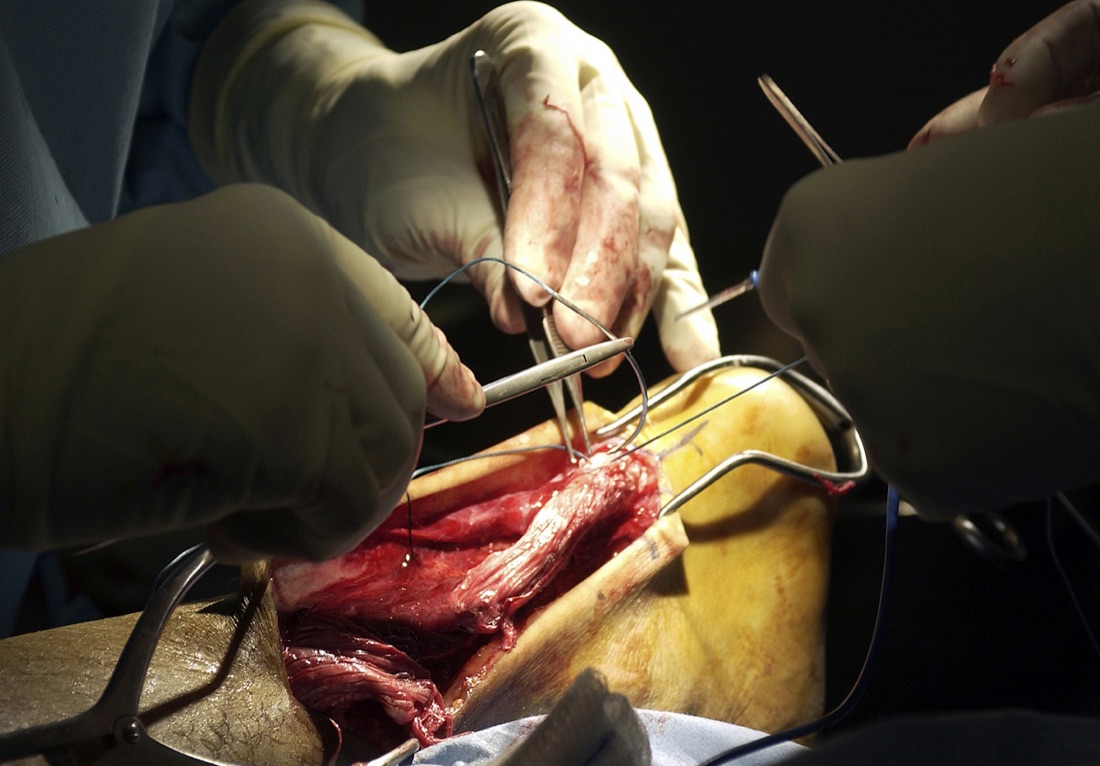
Achilles tendon surgery is performed under spinal/epidural anaesthesia, popliteal nerve block or general anaesthesia. A regional block is effective for pain control in the first 12 - 24 hours after surgery. The patient is usually positioned face down.
General Information About the Operation
Prophylactic antibiotics are given and thromboprophylaxis is planned for the non-weight-bearing period. Skin quality is assessed carefully before surgery — the skin here is thin and its blood supply is limited.
- Duration: Acute repair 45 - 75 minutes; chronic reconstruction 90 - 150 minutes
- Anaesthesia: Spinal/epidural, popliteal block or general anaesthesia
- Hospital stay: Usually day case or one night
- Position: Prone; both legs are compared to set the correct tendon tension
Open Repair — Surgical Technique
- An incision of about 6 - 10 cm is made along the posteromedial border of the tendon; keeping it off the midline reduces shoe pressure and wound problems
- The paratenon is opened carefully and preserved; it is closed separately at the end — important for healing and gliding
- The ruptured ends are identified and frayed tissue is trimmed
- The ends are joined with strong locking suture techniques (Krackow, Bunnell or Kessler type)
- The repair is reinforced circumferentially with fine absorbable sutures
- Tension check: The resting position of both feet is compared on the table; a repair that is too slack or too tight compromises the result
- Paratenon, subcutaneous tissue and skin are closed in layers without tension on the skin
Percutaneous / Minimally Invasive Repair
- Work is done through a small 2 - 3 cm incision at the rupture site and millimetre-sized side entries
- Special guide systems are used to pass sutures through the tendon ends and tie them
- Advantage: Lower risk of wound healing problems and infection than open surgery
- Disadvantage: Higher risk of sural nerve injury; the repair cannot be seen directly
- May be preferred in patients with diabetes, vascular disease or skin problems
Reconstruction for Chronic and Late Ruptures
- V-Y lengthening: A V-shaped incision in the proximal tendon lengthens it; used for gaps of 2 - 5 cm
- Turn-down flap: A strip from the proximal tendon is turned down to bridge the gap
- Flexor hallucis longus (FHL) tendon transfer: The tendon that flexes the big toe is brought down and fixed to the heel bone; preferred for large gaps and poor tissue quality
- Allograft: An option for very large defects
- Healing and return to sport take considerably longer in these cases than after acute repair
Discharge and Weight-bearing
Patients usually go home the same day or the next day. The foot is held pointing downwards (equinus) in a below-knee cast or a walking boot with heel wedges; this position reduces tension on the repair.
The weight-bearing protocol depends on the surgeon. The modern approach, where a strong repair has been achieved, favours early partial weight-bearing in the boot; early loading has a positive effect on tendon healing and muscle strength. Heel height is reduced gradually over 6 - 8 weeks.
Ice for 15 - 20 minutes three to four times a day and elevation of the leg above heart level are recommended. Analgesics, anti-inflammatory medication, stomach protection and anticoagulation until weight-bearing are prescribed. Stitches are removed at 12 - 16 days — later than elsewhere because skin healing is slow in this area.
Early Postoperative Period (First 3 Weeks)
The aims in this period are to secure wound healing, control swelling, protect the repair and maintain thromboprophylaxis. Wound problems are the most common complication of Achilles surgery, so the wound is followed more closely than after other operations.
- Wound care: Discharge, redness, warmth or separation of the wound edges are checked daily. The wound is kept dry for 14 days. Even a small opening in this area can lead to serious problems and should be reported early
- Swelling control: Elevation above heart level, ice and compression stockings
- Maintain equinus: The cast or boot is not removed and the foot is not forced to neutral. The most dangerous movement for the repair is forcing the foot upwards
- Weight-bearing: According to the surgeon's protocol; gradual progression from partial to full weight in the boot
- Early movement (protocol-dependent): Some protocols allow controlled plantar flexion exercises inside the boot from week 2
- Toe, knee and hip exercises: 3 - 4 times a day; reduce swelling and slow muscle wasting
- Thromboprophylaxis: Anticoagulation, compression stockings and toe movement
- Sural nerve monitoring: Numbness along the outer edge of the foot and the little toe should be reported
Follow-up
- Week 1: Wound check
- Days 12 - 16: Removal of stitches
- Week 6: Boot adjustment and weight-bearing decision
- Month 3: Rehabilitation review and strength assessment
- Month 6 and year 1: Return-to-sport decision
Risks & Complications
Achilles tendon surgery is generally successful, but this area has two specific problems: wound healing difficulties because the skin is thin and poorly vascularised, and a risk of re-rupture that exists whichever treatment is chosen.
Risks Specific to Non-operative Treatment
- Re-rupture: Clearly higher than after surgery; usually occurs within the first 3 months, around the time the boot is removed
- Tendon healing elongated: If the gap is large the tendon heals long, causing permanent loss of calf strength and push-off power
- Joint stiffness and muscle wasting from prolonged immobilisation
- Deep vein thrombosis: Risk increases during non-weight-bearing and immobilisation
- More difficult treatment when diagnosis is delayed
Surgical Risks — Common
- Wound healing problems: The most common complication of this operation. The skin is thin and poorly vascularised; wound-edge separation or delayed healing may occur
- Superficial wound infection (usually treated with antibiotics)
- Postoperative pain, swelling and bruising
- Prolonged swelling: May last 6 - 12 months
- Scar sensitivity and discomfort from shoe pressure
- Temporary ankle stiffness — improves with physiotherapy
Surgical Risks — Less Common
- Deep infection: Difficult to treat when it involves the tendon; requires revision, debridement and prolonged antibiotics, sometimes with removal of suture material
- Sural nerve injury: Numbness or a painful neuroma along the outer foot and little toe; more common with percutaneous techniques
- Deep vein thrombosis (DVT) and pulmonary embolism: Increased risk during non-weight-bearing; largely prevented by anticoagulation
- Re-rupture: Markedly lower after surgery than with non-operative treatment, but not zero
- Skin necrosis: Particularly in patients with diabetes, smokers and those with vascular disease; may require a graft or flap
- Repair too tight or too loose: A tight repair limits dorsiflexion; a loose repair reduces push-off power
- Complex regional pain syndrome (CRPS): Rare
Risk Factors
- Smoking: Its adverse effect on wound and tendon healing is particularly marked in this operation
- Poorly controlled diabetes: Markedly increases the risk of wound problems and infection
- Peripheral vascular disease and venous insufficiency
- Corticosteroid use and fluoroquinolone antibiotics
- Advanced age, obesity
- Previous corticosteroid injection into the area
- Delayed diagnosis and treatment
- Poor adherence to the rehabilitation protocol, particularly forcing the foot in the early phase
Contact Your Doctor Immediately If
- There is discharge, a bad smell or an opening in the wound — even a small opening matters in this area
- You have a temperature above 38 °C
- Pain suddenly increases, especially with a 'snapping' sensation during a movement (suspected re-rupture)
- There is firm, one-sided swelling, redness or warmth in the calf (suspected DVT)
- You develop shortness of breath or chest pain (suspected pulmonary embolism)
- Your toes become blue, cold or pale
- Numbness or burning pain along the outer edge of the foot is progressing
- The cast or boot feels excessively tight
Long-term Outcomes
Patient satisfaction is high after correctly treated Achilles tendon ruptures, and most patients return fully to daily activities. Modern functional rehabilitation protocols have markedly narrowed the difference in functional outcome between surgery and non-operative treatment; the main difference lies in the re-rupture rate.
Some loss of calf strength, fewer single-leg heel raises and thinning of the calf may persist for a long time with either treatment. Return to sport usually takes 6 - 12 months; in professional athletes the rate of return to pre-injury performance is limited. Stopping smoking, weight control and adherence to rehabilitation are key determinants of long-term success.
Rehabilitation
Achilles tendon rehabilitation has to balance two opposing needs: not stressing the repair in the early phase, and moving as early as possible to prevent muscle wasting and adhesions. Modern functional protocols achieve this balance by protecting the foot in equinus while allowing controlled movement and early weight-bearing.
Rehabilitation After Surgery
- Weeks 0 - 2: Foot in equinus in a cast or heel-wedged boot. Elevation, ice and swelling control. Toe, knee and hip exercises. Mobilising with crutches; partial weight-bearing may be allowed according to the surgeon's protocol.
- Weeks 2 - 6: Heel height is gradually reduced. Controlled active plantar flexion and gentle dorsiflexion begin in the boot (the foot is not forced beyond neutral). Progression from partial to full weight-bearing. Scar massage once the wound has healed.
- Weeks 6 - 10: Heel wedges are removed and the foot reaches neutral. Gradual transition from the boot to a normal shoe (with heel support). Ankle range-of-movement work intensifies. Stationary cycling can begin.
- Months 3 - 4: Calf strengthening takes priority. Begin with double-leg heel raises and progress gradually to single-leg. Balance and proprioception exercises. Swimming and pool walking.
- Months 4 - 6: Eccentric strengthening (controlled heel drops from a step) is added — the most important exercise for tendon healing. Light running on flat ground can begin if criteria are met.
- Months 6 - 9: Gradual introduction of sport-specific drills; jumping and change-of-direction work. Plyometric exercises.
- Months 9 - 12: Full return to contact and high-intensity sport once criteria are met and with the surgeon's approval.
Rehabilitation With Non-operative Treatment
- Weeks 0 - 2: Foot in equinus in a cast or boot; ultrasound confirms the ends are apposed. Elevation, ice, thromboprophylaxis.
- Weeks 2 - 8: Heel height reduced gradually; controlled movement and progressive weight-bearing in the boot. A functional protocol is clearly superior to complete immobilisation.
- Weeks 8 - 12: Transition from boot to shoe; range of movement and light strengthening.
- Months 3 - 6: Calf strengthening, balance and proprioception. Eccentric programme.
- Months 6 - 12: Gradual return to sport. Because the risk of re-rupture is highest in the first 3 months, adherence to the protocol in this period is critical.
Return-to-sport Criteria
- Pain-free, near-symmetrical ankle range of movement
- Single-leg heel raise repetitions at least 85 - 90% of the other leg
- Difference in calf circumference less than 2 cm
- 90% symmetry on single-leg hop tests
- Pain-free running on flat ground without a limp
- Pain-free completion of sport-specific movements
- No swelling or pain after activity
- Approval from the surgeon and physiotherapist
Recommended Activities
- Stationary cycling and elliptical trainer (non-weight-bearing cardiovascular activity in the early phase)
- Swimming and pool walking (once the wound has healed)
- Walking — distance increased gradually
- Eccentric calf exercises (according to protocol, from month 4)
- Balance board and proprioception work
Avoid in the First 6 Months
- Sudden acceleration, jumping and changes of direction
- Running quickly up or down stairs and slopes
- Situations that could force the foot suddenly upwards (missing a step)
- Running on uneven ground
- Sports requiring explosive push-off such as tennis, basketball and football
- Long walks in flat, unsupportive shoes
Lifestyle and Recovery Advice
- Do your home exercise programme every day; eccentric exercises in particular determine the quality of tendon healing
- Do not smoke — its adverse effect on wound and tendon healing is particularly marked in Achilles surgery
- If you have diabetes, keep your blood glucose tightly controlled
- Healthy weight: Every extra kilogram increases the load on the Achilles tendon
- Follow the weight-bearing and boot programme strictly; forcing the foot early is the most common cause of re-rupture
- Keep the leg above heart level as much as possible in the first months
- Footwear: In the first months choose shoes with heel support and a soft back
- Take care of the other Achilles tendon: The risk is increased on the other side after a rupture
- If you are prescribed a fluoroquinolone antibiotic, tell your doctor about your Achilles rupture
- Attend regular follow-up (week 6, month 3, month 6, year 1)
What to Expect
Walking usually normalises by month 3 and return to daily activities is complete at 3 - 4 months. Running resumes at 4 - 6 months and full return to sport at 6 - 12 months. Full functional recovery may take 12 - 18 months.
Some persistent loss of calf strength and thinning of the calf is expected; disciplined eccentric strengthening narrows this difference considerably. Mild swelling and stiffness after activity may last 6 - 12 months. Patient, consistent rehabilitation is the single most important factor in the outcome of this injury.
Is This Treatment Right for You?
Every patient's condition is different. After a detailed examination and imaging, the treatment plan that suits you best will be prepared.
Would you like more information about Achilles Tendon Rupture?
Book Appointment